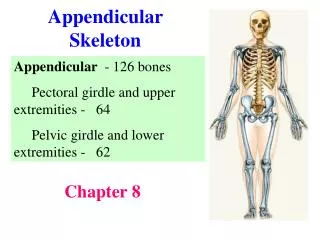
Appendicular Skeleton: Pectoral Girdle and Upper Limbs
Learn about the pectoral girdle and upper limbs, including the bones of the shoulder, arm, forearm, wrist, palm, and fingers. Discover their surface markings and common disorders.

Appendicular Skeleton: Pectoral Girdle and Upper Limbs
E N D
Presentation Transcript
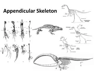
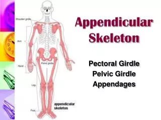
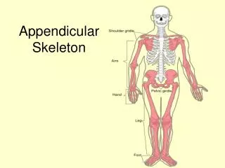
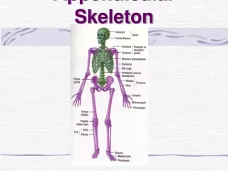
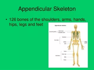
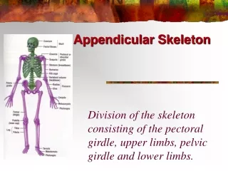
Appendicular Skeleton Division of the skeleton consisting of the pectoral girdle, upper limbs, pelvic girdle and lower limbs.
Pectoral Girdle • Attaches the bones of the upper limbs to the axial skeleton
Clavicle • Also known as the collarbone • Long, slender S-shaped bone that lies horizontally above the first rib (Transmits mechanical force from the upper limb to the trunk)
Scapula • Also known as the shoulder blade • Large, flat triangular bone on the posterior part of the thorax
SPINE: A sharp ridge that runs diagonally across the back portion of the scapula body • BODY – Main flat area of the scapula • ACROMION: The lateral end of the spine. Where the scapula articulates with the clavicle
GLENOID CAVITY (glenoid fossa) - a depression inferior to the acromion where the head of the humerus sits • CORACOID PROCESS – Projection anterior to the acromion for muscle attachment
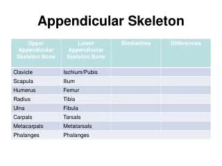
Upper Limb • Consists of 30 bones (all paired up) • Humerus in the arm • Ulna and radius in the forearm • 8 carpals, 5 metacarpals, and 14 phalanges in the hand
Humerus • Longest and largest bone of the upper limb • Articulates with the scapula at the shoulder and both the ulna and radius at the elbow
Humerus Bone Surface Markings • ANATOMICAL NECK: constricted portion distal to the head – site of the epiphyseal plate
BODY: Main portion of the bone (diaphysis) • DELTOID TUBEROSITY: a roughened V-shaped area where the deltoid muscle attaches
CAPITULUM – small rounded process at the distal end that articulates with the head of the radius. • RADIAL FOSSA - a depression that receives the head of the radius when the forearm is bent.
TROCHLEA - a spool-shaped surface that articulates with the ulna. • CORONOID FOSSA – a depression that receives part of the ulna when the forearm is bent. • OLECRANON FOSSA - a depression on the back of the bone that receives the ulna when the forearm is straightened.
Ulna • Located on the medial side of the forearm (pinky side) • Longer than the radius
Ulna Bone Surface Markings • The olecranon forms the prominence of the elbow on the proximal end. • The coronoid process projection on the proximal, helps to hold the trochlea • Trochlear Notch – depression formed by the olecranon and coronoid process
The radial notch is a depression for the head of the radius. • A styloid process is a pointy projection at the distal end.
Radius • Located on the lateral side of the forearm (thumb side)
Radius Bone Surface Markings • Radial tuberosity a raised, roughened area that is where the biceps brachii muscle attaches to the bone • Styloid Process – pointy projection on the distal end
Carpus (Wrist) • 8 carpals • Held together by ligaments with four bones in each row • Named for their shapes • Short bones
The carpals in the proximal (closest to the radius/ulna) row are the: • Scaphoid, Lunate, Triquetrum, and Pisiform • The carpals in the distal row are the: • Trapezium, Trapezoid, Capitate, and Hamate
Metacarpus (Palm) • 5 metacarpals • Each consists of a proximal base, an intermediate body, and a distal head • Numbered I-V starting with the thumb • Long bones
Phalanges (Fingers) • 14 in each hand • Thumb has two (proximal and distal) • In each of the other four digits, there are three (proximal, middle, and distal)
Carpal Tunnel Syndrome • Narrowing of the carpal tunnel causes compression of the median nerve • The nerve compression causes pain, numbness, tingling, and hand muscle weakness
Rotator Cuff Injury • Tears or inflammation of ligaments and tendons of the shoulder near the humerus • Results in pain and loss of shoulder mobility
Checkpoint Questions • Which bones make up a pectoral girdle? What is the function of the pectoral girdle? • With which part of the scapula does the humerus articulate? • What part of the ulna is called the “elbow”? • What part of which bones are commonly called the “knuckles”? • What bones form the upper limb, from proximal to distal?
Pelvic (hip) Girdle • Functions: • Support for vertebral column • Protect pelvic organs • Attach lower limbs • Coxal Bones: Hip bones • 3 parts: Pubis, Ilium and Ischium
Articulations • Sacroiliac Joint – posterior articulation of the pelvic girdle • Pubic Symphysis – anterior articulation of the pelvic girdle • Acetabulum – attachment point of the femur • socket of the ball and socket joint
Coxal Bones • Pubis – anterior portion • Joined by pubic symphysis • Ilium – superior portion • Iliac Crest – ridge at the top of the ilium • Ischium – inferior portion • Acetabulum – socket for the head of the femur • Obturator Foramen – hole formed by the ischium and pubis
Pelvis • Combination of the sacrum, coccyx, and the 2 hip bones • Greater (false) – Top portion that is not fully enclosed by bone • Lesser (true) – Bottom portion the is completely surrounded by bone
Pelvis - continued • Ilium • Ischium • Pubis • Pubic Symphysis • Sacrum • Coccyx • Pelvic Brim • Pubic Arch
Pelvimetry • Measurement of the size of the inlet and outlet of the birth canal.
Pelvic Girdle Checkpoint • What is the name of the hip bone? • What are the 3 parts of the hip bone? • Can you identify them on a diagram??? • List 3 functions of the pelvic girdle • What is the name of the “socket” where the head of the femur sits? • List 3 differences between the male and female pelvis. Why are these present?
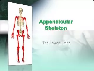
LOWER LIMB • Includes the thigh, leg, ankle, foot and toes • 30 bones in each • Femur • Patella • Tibia • Fibula • Tarsals • Metatarsals • Phalanges
Femur • Thigh Bone • Longest, strongest heaviest bone in the body • Diaphysis has a medial bend to bring knees closer to the midline of the body
Femur continued • Body - diaphysis • Head – “ball” of ball and socket joint • Neck – common site of fractures • Greater and Lesser Trochanters – used for muscle attachment • Lateral and Medial Condyles – articulation with the tibia • Patellar Surface
Patella • Sesamoid bone • Develops in the tendon of the quadriceps femoris muscle • Increases the leverage of the tendon and maintains the position of the tendon
Patellofemoral stress syndrome • AKA “Runner’s Knee” • Patella does not glide up and down between the femoral condyles but rather laterally causing pain.