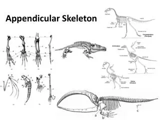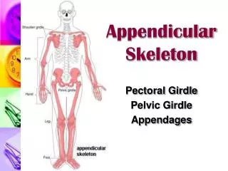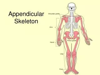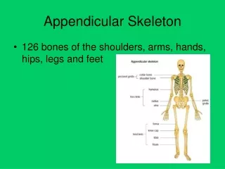
APPENDICULAR SKELETON
APPENDICULAR SKELETON. CHAPTER 8. THE PECTORAL GIRDLE. SECTION IV. Pectoral or Shoulder Girdle. Consists of two bones, the anteriorly positioned clavicle and the posteriorly positioned scapula

APPENDICULAR SKELETON
E N D
Presentation Transcript
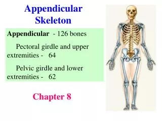
APPENDICULAR SKELETON CHAPTER 8
THE PECTORAL GIRDLE SECTION IV
Pectoral or Shoulder Girdle • Consists of two bones, the anteriorly positioned clavicle and the posteriorly positioned scapula • Pectoral girdle is a loosely attached, held in place largely by musculature attached to the thorax and the vertebral column • Only direct ligament attachment exists at the sternoclavicular joint • Frees girdle to move over the thorax as the need arises
Flexible and Mobile • Pectoral girdle is very light to allow the upper limb flexibility and mobility not allowed anywhere else in body • This is possible because only the sternal end of clavicle is attached to axial skeleton thus allowing the scapula to move across thorax and the arm with it • The socket of the shoulder joint is shallow and poorly reinforced • Although this arrangement does not restrict movement it is less stable
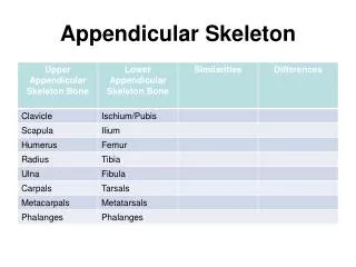
Clavicles • Clavicles are double curved bones extending along the superior thorax • Attached on the sternal end to sternum and the acromial • Attachment site or muscles of the thorax and shoulder • Position scapula away from thorax
Right ScapulaAnterior Aspect • Bone markings are related to • Joint structures • Muscle attachments • Nerve and blood vessels
Right ScapulaPosterior Aspect • Bone markings are related to • Joint structures • Muscle attachments • Nerve and blood vessels
Right ScapulaLateral Aspect • Schematic representation of its orientation
THE UPPER LIMB SECTION V
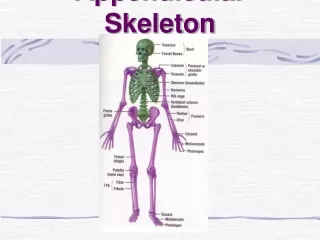
The Upper Limb • Arm • Humerus • Forearm • Ulna • Radius • Hand • Wrist (8 carpal bones) • Palm (5 metacarpal bones) • Fingers (14 phalanges)
Radius and ulna • Ulna is involved in elbow flexion • Radius is involved with supination and pronation
THE PELVIC GIRDLE SECTION VI
The Pelvic (Hip) Girdle • Attaches the lower limbs to axial skeleton • Transfers the weight of the torso, head, and upper extremities to lower limbs • Supports the visceral organs of the pelvis • Secured by strong ligaments and deep sockets the joint is reinforced for stability • Less range of motion in all planes of movement • Female pelvic structure to facilitate childbearing
Pelvic girdle is formed by a pair of coxal bones, each called an os coxae Each os coxae unites anteriorly at the pubic symphysis and with the sacrum posteriorly Each coxa is formed by the ilium, ischium and pubic which were separate during childhood but fused in adulthood Collectively the os coxae, sacrum and coccyx is called the pelvis Pelvis
Os Coxa • Ilium • Superior • Ishium • Posterior • Pubis • Anterior
Os Coxa • Ilium • Superior • Ishium • Posterior • Pubis • Anterior
Pelvic Structure and Childbearing • The female pelvis reflects modifications for child bearing • It tends to be wider, shallower, lighter, and rounder than the male • Pelvic modifications accommodate the growing fetus as well as providing a birth canal wide enough to allow the infants head to exit at birth • Pelvic inlet and outlet are critical to delivery
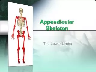
THE LOWER LIMB SECTION VII
The Lower Limb • Thigh • Femur • Leg • Tibia • Fibula • Foot (7 Tarsal bones) • Instep (5 Metatarsal bones) • Toes (14 Phalanges)
Tibia Fibula
Bones of Right Foot
DEVELOPMENTAL ASPECT OF THE SKELETON SECTION VIII
Developmental Aspects of the Skeleton • Fontanels • Spinal Curvatures • Long Bone Ratio • Changes in Female Pelvis • Adult Skeletal Changes