Download

1 / 46
490 likes | 1.37k Views
Learn advanced airway management techniques, including anatomy, equipment use, intubation skills, and patient care considerations. This guide covers gastric tubes, Sellick maneuver, endotracheal intubation, and more.

E N D
Cognitive Objectives (1 of 5) 8-1.1 Identify and describe the airway anatomy in the infant, child, and the adult. 8-1.3 Explain the pathophysiology of airway compromise. 8-1.4 Describe the proper use of airway adjuncts. 8-1.5 Review the use of oxygen therapy in airway management.
Cognitive Objectives (2 of 5) 8-1.6 Describe the indications, contraindications, and techniques for insertion of nasal gastric tubes. 8-1.7 Describe how to perform the Sellick maneuver (cricoid pressure). 8-1.8 Describe the indications for advanced airway management.
Cognitive Objectives (3 of 5) 8-1.9 List the equipment required for orotracheal intubation. 8-1.10 Describe the proper use of the curved blade for orotracheal intubation. 8-1.11 Describe the proper use of the straight blade for orotracheal intubation. 8-1.12 State the reasons for and proper use of the stylet for orotracheal intubation.
Cognitive Objectives (4 of 5) 8-1.13 Describe the methods of choosing the appropriate size endotracheal tube in an adult patient. 8-1.14 State the formula for sizing an infant or child endotracheal tube. 8-1.15 List complications associated with advanced airway management. 8-1.17 Describe the skill of orotracheal intubation in the adult patient.
Cognitive Objectives (5 of 5) 8-1.18 Describe the skill of orotracheal intubation in the infant and child patient. 8-1.19 Describe the skill of confirming endotracheal tube placement in the adult, infant, and child patient. 8-1.20 State the consequences of and the need to recognize unintentional esophageal intubation. 8-1.21 Describe the skill of securing the endotracheal tube in the adult, infant, and child patient.
Affective Objectives (1 of 2) 8-1.22 Recognize and respect the feelings of the patient and family during advanced airway procedures. 8-1.23 Explain the value of performing advanced airway procedures. 8-1.24 Defend the need for the EMT-B to perform advanced airway procedures. 8-1.25 Explain the rationale for the use of a stylet.
Affective Objectives (2 of 2) 8-1.26 Explain the rationale for having a suction unit immediately available during intubation attempts. 8-1.27 Explain the rationale for confirming breath sounds. 8-1.28 Explain the rationale for securing the endotracheal tube.
Psychomotor Objectives 8-1.29 Demonstrate how to perform the Sellick maneuver. 8-1.30 Demonstrate the skill of orotracheal intubation in the adult patient. 8-1.31 Demonstrate the skill of orotracheal intubation in the infant and child patient. 8-1.32 Demonstrate the skill of confirming endotracheal tube placement in the adult patient. 8-1.33 Demonstrate the skill of confirming endotracheal tube placement in the infant and child patient. 8-1.34 Demonstrate the skill of securing the endotracheal tube in the adult patient.
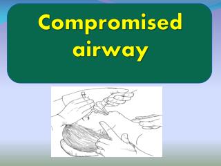
Basic Airway Management • Airway is always assessed first. • Advanced techniques are used after basic management. • The first step is opening the patient’s airway. • Once the airway has been cleared, determine the need for an airway adjunct.
Gastric Tubes • Provide channel into patient’s stomach • Nasogastric tubes: Inserted through the nose • Orogastric tubes: Inserted through the mouth • Nasogastric tubes: Contraindicated in a patient with major facial, head, or spinal trauma
Equipment • Proper-sized tubes • Catheter-tipped 60-mL syringe • Water-soluble lubricant • Emesis container • Tape • Stethoscope • Suctioning unit and catheters
Measure the tube. Lubricate the distal end of the tube. Place the patient in proper position. Pass the tube until you reach the tape marker. Confirm proper tube placement. Aspirate air and stomach contents with the syringe. Secure the tube in place with tape. Gastric Tube Insertion
Sellick Maneuver • Visualize the cricoid cartilage. • Palpate to confirm its location. • Apply firm pressure on the cricoid ring. • Maintain pressure until intubated.
Endotracheal Intubation • Insertion of a tube into the trachea in order to maintain the airway • Orotracheal intubation: Through the mouth • Nasotracheal intubation: Through the nose • EMT-Bs only intubate patients who are: • Unresponsive with no gag reflex • In cardiac arrest
Equipment (1 of 2) • BSI equipment • Proper-equipment endotracheal tube (ET tube) • Laryngoscope handle and blade (visualized technique) • Stylet or light stylet • 10-mL syringe • Oxygen, with BVM device
Equipment (2 of 2) • A suctioning unit with rigid and soft-tip catheters • Magill forceps • Towels for raising the patient’s head and/or shoulders • A stethoscope • Water-soluble lubricant for tubes and scopes • A commercial securing device or tape
Laryngoscope • Sweeps the tongue out of the way and aligns the airway • Has a light powered by batteries in handle • Has blades that connect to handle • Blades are curved or straight. • They range in size from 0 to 4.
Endotracheal Tubes • Tubes come in many sizes, from adult to infant. • Normal tube-to-teeth mark is usually around 22 cm. • Diameter for normal adult male ranges from 7.5 to 8.5 mm. • Diameter for normal adult female ranges from 6.5 to 8.0 mm. • Use tape or chart for pediatric sizes.
Stylet • Plastic-coated wire may be inserted in the ET tube to add rigidity and shape to the tube. • Bend the tip of the stylet to form a gentle curve in adults. • Bend the tip of the stylet to form a hockey stick shape for an infant and child. • Confirm that the stylet is not sticking out past the end of the ET tube.
Syringe • Use the 10-mL syringe to test for air leaks in the ET tube before intubation. • After the ET tube has been properly inserted, inflate the cuff with 5 to 10 mL of air. • Remove the syringe from the pilot balloon to prevent air from leaking.
Other Equipment • Oxygen • A suctioning unit • A BVM device • Magill forceps • Towels for raising the patient’s head or shoulders • Secondary confirmation device • C-collar backboard
The Intubation Procedure • First EMT-B applies AED. • Second and third EMT-B perform CPR. • Fourth EMT-B prepares and intubates patient.
Visualized (Oral) Intubation (1 of 2) • Open airway. • Insert an oropharyngeal airway. • Preoxygenate the patient. • Assemble equipment. • Position the head and neck.
Visualized (Oral)Intubation (2 of 2) • Grasp laryngoscope with left hand. • Visualize vocal cords. • Insert ET tube. • Inflate balloon. • Confirm placement. • Secure tube.
Blind (Nasal) Intubation (1 of 2) • Many of the steps are the same as those for oral intubations. • Preoxygenate the patient. • Check for gag reflex. • Insert tube through nostril. • Pass tube through vocal cords as patient is inhaling.
Blind (Nasal) Intubation (2 of 2) • Release the jaw and hold tube against nostril. • Inflate cuff. • Attach the BVM device. • Confirm placement. • Secure the tube.
Intubating the right main stem bronchus Intubating the esophagus Aggravating spinal injuries Taking too long to ventilate Patient vomiting Soft-tissue trauma Mechanical failure Patient intolerant of the ET tube Decrease in heart rate Intubation Complications
Multilumen Airways • Inserted without direct visualization • Provide ventilation when placed in either trachea or esophagus
Combitube Contraindications • Conscious or semiconscious patients with gag reflex • Children younger than 16 years • Adults shorter than 5' • Patients who have ingested a caustic substance • Patients with esophageal disease
Inserting the ETC (1 of 2) • Assemble and check the proper equipment. • Apply water-soluble lubricant to the ETC. • Position the patient. • Preoxygenate the patient. • Lift the lower jaw and tongue.
Inserting the ETC (2 of 2) • Guide the ETC along the base of the tongue. • Inflate the blue and then the white pilot balloon. • Ventilate the patient. • Confirm placement. • Monitor the patient.
Removing the ETC • Be prepared to suction patient. • Deflate both balloon cuffs. • Gently remove the tube.
PtL Contraindications • Conscious or semiconscious patients with gag reflex • Children younger than 14 years • Adults shorter than 5' • Patients who have ingested a caustic substance • Patients with esophageal disease
Inserting the PtL (1 of 2) • Assemble and check equipment. • Lubricate tube with water-soluble lubricant. • Position the patient. • Preoxygenate the patient. • Lift the lower jaw and tongue. • Hold the PtL so that it curves in the same direction as the pharynx.
Inserting the PtL (2 of 2) • Inflate balloon cuffs. • Ventilate patient through the short, green tube. • Evaluate placement. • Verify that the patient is receiving adequate ventilations. • Monitor the patient.
Removing the PtL • Be prepared to suction the patient. • Deflate balloon cuffs. • Gently remove the tube.
LMA Contraindications • Asthma • COPD • Leaking mask • Active vomiting • Esophageal diseases
Inserting the LMA (1 of 2) • Assemble and check equipment. • Open the airway. • Preoxygenate the patient. • Select proper size. • Hold LMA down. • Remove oropharyngeal device and begin insertion.
Inserting the LMA (2 of 2) • Insert until you feel resistance. • Stabilize the tube. • Inflate mask. • Confirm placement. • Insert bite block and secure the LMA.