Download
1 / 40
410 likes | 620 Views
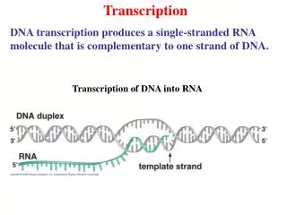
Transcription. Chapter 11. 11.1 Introduction. Figure 11.1. Figure 11.2. 11.2 Transcription Occurs by Base Pairing in a “Bubble” of Unpaired DNA. RNA polymerase separates the two strands of DNA in a transient “bubble.”

E N D
Transcription Chapter 11
11.1 Introduction Figure 11.1 Figure 11.2
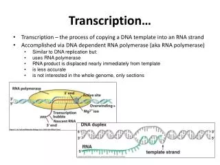
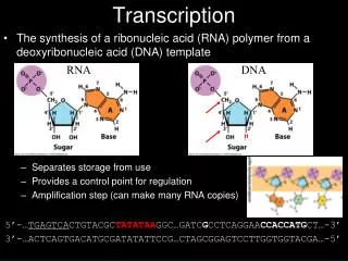
11.2 Transcription Occurs by Base Pairing in a “Bubble” of Unpaired DNA • RNA polymerase separates the two strands of DNA in a transient “bubble.” • It uses one strand as a template to direct synthesis of a complementary sequence of RNA. • The length of the bubble is ∼12 to 14 bp • The length of RNA-DNA hybrid within it is ∼8 to 9 bp. Figure 11.3
11.3 The Transcription Reaction Has Three Stages • RNA polymerase initiates transcription after binding to a promoter site on DNA. • During elongation the transcription bubble moves along DNA. • The RNA chain is extended in the 5′–3′ direction. • When transcription stops: • the DNA duplex reforms • RNA polymerase dissociates at a terminator site Figure 11.6
11.4 Phage T7 RNA Polymerase Is a Useful Model System • T3 and T7 phage RNA polymerases are single polypeptides. • They have minimal activities in recognizing a small number of phage promoters. • Crystal structures of T7 RNA polymerase with DNA identify: • the DNA-binding region • the active site Figure 11.7
11.5 A Model for Enzyme Movement Is Suggested by the Crystal Structure • DNA moves through a groove in yeast RNA polymerase that makes a sharp turn at the active site. Figure 11.12
A protein bridge changes conformation to control the entry of nucleotides to the active site. Figure 11.15
11.6 Bacterial RNA Polymerase Consists of Multiple Subunits • Bacterial RNA core polymerases are ∼500 kD multisubunit complexes with the general structure α2ββ′. Figure 11.16
DNA is bound in a channel and is contacted by both the β and β′ subunits. Figure 11.17
11.7 RNA Polymerase Consists of the Core Enzyme and Sigma Factor • Bacterial RNA polymerase can be divided into: • the α2ββ′ core enzyme that catalyzes transcription • the sigma subunit that is required only for initiation
Sigma factor changes the DNA-binding properties of RNA polymerase: • its affinity for general DNA is reduced • its affinity for promoters is increased Figure 11.18
Binding constants of RNA polymerase for different promoters vary over six orders of magnitude. • The variation corresponds to the frequency with which transcription is initiated at each promoter.
11.8 The Association with Sigma Factor Changes at Initiation • When RNA polymerase binds to a promoter, it: • separates the DNA strands to form a transcription bubble • incorporates up to nine nucleotides into RNA • There may be a cycle of abortive initiations before the enzyme moves to the next phase. • Sigma factor may be released from RNA polymerase when the nascent RNA chain reaches eight to nine bases in length.
11.9 A Stalled RNA Polymerase Can Restart • An arrested RNA polymerase can restart transcription by cleaving the RNA transcript to generate a new 3′ end.
11.10 How Does RNA Polymerase Find Promoter Sequences? • The rate at which RNA polymerase binds to promoters is too fast to be accounted for by random diffusion. Figure 11.22
RNA polymerase probably: • binds to random sites on DNA • exchanges them with other sequences very rapidly until a promoter is found Figure 11.23
11.11 Sigma Factor Controls Binding to DNA • A change in association between sigma factor and holoenzyme changes binding affinity for DNA. • This allows core enzyme to move along DNA. Figure 11.25
11.12 Promoter Recognition Depends On Consensus Sequences • A promoter is defined by the presence of short consensus sequences at specific locations.
The promoter consensus sequences consist of: • a purine at the startpoint • the hexamer TATAAT centered at –10 • another hexamer centered at –35 • Individual promoters usually differ from the consensus at one or more positions. Figure 11.26
11.13 Promoter Efficiencies Can Be Increased or Decreased by Mutation • Down mutations to decrease promoter efficiency usually decrease conformance to the consensus sequences. • Up mutations have the opposite effect. • Mutations in the –35 sequence usually affect initial binding of RNA polymerase.
Mutations in the –10 sequence usually affect the melting reaction that converts a closed to an open complex. Figure 11.27
11.14 RNA Polymerase Binds to One Face of DNA • The consensus sequences at –35 and –10 provide most of the contact points for RNA polymerase in the promoter. • The points of contact lie on one face of the DNA. Figure 11.29
11.15 Supercoiling Is an Important Featureof Transcription • Negative supercoiling increases the efficiency of some promoters by assisting the melting reaction. • Transcription generates: • positive supercoils ahead of the enzyme • negative supercoils behind it • These must be removed by gyrase and topoisomerase. Figure 11.30
11.16 Substitution of Sigma Factors MayControl Initiation • E. coli has several sigma factors. • Each causes RNA polymerase to initiate at a set of promoters defined by specific –35 and –10 sequences. Figure 11.31
σ70 is used for general transcription • The other sigma factors are activated by special conditions. Figure 11.34
11.17 Sigma Factors Directly Contact DNA • σ70 changes its structure to release its DNA-binding regions when it associates with core enzyme. • σ70 binds both the –35 and –10 sequences. Figure 11.37
11.18 Sigma Factors May Be Organized intoCascades • A cascade of sigma factors is created when one sigma factor is required to transcribe the gene coding for the next sigma factor.
The early genes of phage SPO1 are transcribed by host RNA polymerase. • One of the early genes codes for a sigma factor that causes RNA polymerase to transcribe the middle genes. • Two of the middle genes code for subunits of a sigma factor that causes RNA polymerase to transcribe the late genes. Figure 11.40
11.19 Sporulation Is Controlled by Sigma Factors • Sporulation divides a bacterium into a mother cell that is lysed and a spore that is released. Figure 11.41
Each compartment advances to the next stage of development by synthesizing a new sigma factor. • The new sigma factor displaces the previous one. Figure 11.42
Communication between the two compartments coordinates the timing of sigma factor substitutions. Figure 11.43
11.20 Bacterial RNA Polymerase Terminates at Discrete Sites • Termination may require both: • recognition of the terminator sequence in DNA • formation of a hairpin structure in the RNA product Figure 11.45
11.21 There Are Two Types of Terminators in E. coli • Intrinsic terminators consist of: • a G-C-rich hairpin in the RNA product • followed by a U-rich region in which termination occurs Figure 11.46
11.22 How Does Rho Factor Work? • Rho factor is a terminator protein that: • binds to a rut site on nascent RNA • tracks along the RNA to release it from the RNA–DNA hybrid structure at the RNA polymerase Figure 11.47
11.23 Antitermination Is a Regulatory Event • Termination is prevented when antitermination proteins act on RNA polymerase. • This causes it to read through a specific terminator or terminators. Figure 11.51
Phage lambda has two antitermination proteins, pN and pQ. • They act on different transcription units. Figure 11.52
11.24 Antitermination Requires Sites That Are Independent of the Terminators • The site where an antiterminator protein acts is upstream of the terminator site in the transcription unit. • The location of the antiterminator site varies in different cases and can be: • in the promoter or • within the transcription unit Figure 11.53
11.25 Termination and AntiTermination Factors Interact with RNA Polymerase • Several bacterial proteins are required for lambda pN to interact with RNA polymerase. Figure 11.55
These proteins are also involved in antitermination in the rrn operons of the host bacterium. • The lambda antiterminator pQ has a different mode of interaction that involves binding to DNA at the promoter.