Download

1 / 64
640 likes | 819 Views
Breast Diseases. Breast diseases. Anatomy Physical Examination Acute Mastitis Cystic hyperplasia Breast Tumor Gynecomastia. Anatomy. On pectoral fascia and musculature of the chest wall Over upper anterior rib cage 2 nd or 3 rd to 6 th Fat surrounding Skin envelope

E N D
Breast diseases • Anatomy • Physical Examination • Acute Mastitis • Cystic hyperplasia • Breast Tumor • Gynecomastia
Anatomy • On pectoral fascia and musculature of the chest wall • Over upper anterior rib cage 2nd or 3rd to 6th • Fat surrounding • Skin envelope • Axillary tail of Spencer
Anatomy • 15-20 glandular lobes • 20-40 lobules • 10-100 alveoli • Small duct-major duct • Nipple-areolar complex • Cooper’s ligament (fibrous septa)
Anatomy • Relation to pectoralis major muscle • Level I Encompasses the LN lateral to lateral border of the pectoralis minor muscle; this subgroup contains most of the axillary nodes.
Anatomy • Level II LN lying directly beneath the pectoralis minor muscle • Level III Medial to the medial border of the pectoralis minor muscle and extending up to the apex of the axilla
lymph • Pectoralis major → axillary → subclavicular → supra-clavicular • Medial portion → intercostal lymphatic duct →para-mediastinum
Lymph • Subcutaneous lymphatic communication left→right • Lymphatic plexus on the rectal sheath • →falciform ligament→liver
Important structures • Intercostobrachial nerve • a sensory nerve supplying the underarm skin • Long thoracic nerve of Bell • a motor nerve to the serratus anterior • and subscapularis muscles • Thoracodorsal nerve • a motor nerve to the latissimus dorsi adjacent • to its accompanying artery and veins
Physiology • Anterior pituitary hormones (prolactin) • Adrenal corticoid hormones (Estrogen) • Sexual hormones (Progesterone) • Insulin • Thyroid hormone
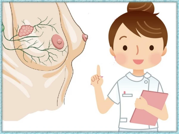
Examination Inspection • Overall inspection symmetry, size, shape, skin color, venous pattern, lump, local dimpling • Nipple excoriation, inversion, discharge, edema and redness • Skin redness, edema, Peau d’orange or pig-skin
Examination Palpation • Gentle palpation, quadrant by quadrant • Mass: number, size, consistency and mobility • Lymph node: Central,pectoral,subscapular , subclavicular and supra-clavicular group
Examination palpation • Character of the discharge is significant Milky, serous, or green-brown discharge Bloody discharge
Imaging Study • Mammography • Thermography • Ultrasound • Ductogram • Magnetic Resonance (MR) • Positron Emission Tomography (PET)
Evaluation of breast masses • Biopsy : FNA(0.7-0.9mm) Open biopsy • Nipple discharge: serous,colorless • Normal menstrual cycle, intraductal papilloma or early pregnancy • Bloody intraductal papilloma or ductal ca. • Yellowish galactocele or cystic hyperplasia • Ductogram
Acute Mastitis • Cause Lactic stasis Bacterial invasion (Staphylococcus aureus) • Manifestation Swelling pain Painful mass with reddish skin General features: Chill, fever, ipsilateral LN enlargement, bacteriaemia Abscess formation
Treatment • General • Thermo therapy: 25% Magnesium sulfate • Antibiotic therapy: Local and general administration • Drainage: • Prevention
Cystic hyperplasia Cause • Hormonal imbalance • Excessive estrogen production and deficient corpus luteinum activity
Cystic hyperplasia Clinical manifestation • Pain or lump, nipple discharge (15%) • Bilateral invlvement • Inseparable from other grandular tissue • Tense cyst no fluctuant Tenderness • Cyst may appear rapidly and then maintain their size or shrink after next menstraual flow • Most painful in pre-menstraual period: mastodynia
Cystic hyperplasia Diagnosis • Pain or lump • FNA Management Hormonal therapy FNA, open biopsy, mastectomy
Tumor • Benign Fibroadenoma 75% Intraductal papiloma 20% • Malignant Cacer 98% Sarcoma 2%
Fibroadenoma Cause Estrogen may play an important role in its pathogenesis
Fibroadenoma Manifestation Lump or mass Freely moveable, smooth, lobulated and independent from surroundings without fixation regardless the size. 75% solitary Found incidentally on BSE
Fibroadenoma Diagnosis Mammographic appearance of a degenerating firoadenoma displaying a characteristic pattern of dense, popcorn-like calcification Histological appearance of a typical firoadenoma
Fibroadenoma Treatment Excision continued growth and need to be certain of the diagnosis
Intraductal papilloma • Woman of 40-50 years old. • 6%-8% malignant tendency. • Forming from the epithelial linings of the main ducts.
Intraductal papilloma • Nodule at the areola margin. • Pressure at that point reproduces the bloody discharge. • Surgical excision (involved duct or radical resection if it is proved malignant by frozen section)
Breast Cancer Every 12 minutes a woman in America dies ofbreast cancer
CHANGES IN BREAST CANCER SURGERY, 1972–1986 • “LESS CAN BE BETTER”
Breast cancer for women • Cancer No.1or 2 for • Death No.5 • Shanghai Morbidity: in 1972 18.90/100,000 in 1999 52.98/100,000
Etiology • Estrogen as an important factor in pathogenesis Estron(E1) and estradiol (E2) : carcinogenic Estriol (E3) : non-carcinogenic • Other various factors
Etiology • Cumulative risk of developing invasive beast cancer after a biopsy for benign breast disease • Women with proliferative disease with atypia are at significantly increased risk for developing invasive breast cancer
Etiology • Age-specific incidence curves for breast cancer • The curve rises sharply after 30 years of age and continues to climb thereafter
Demographic factor • Age more than 30 yrs • Female gender (130:1 female/male ratio) • Greatly increased risk • Known carrier of breast cancer susceptibility gene • Strong family history—2 or more first-degree relatives with bilateral or premenopausal breast cancer • Atypical ductal or lobular hyperplasia or lobular carcinoma in situ • Ductal carcinoma in situ, risk limited to ipslateral breast
Pathological procedure • Tumor size • Hormone receptor status • Status of excision margins • Histologic type
Pathological classification Type 1 Non-metastasizing Inarticulate carcinoma Type 2 Rarely metastasizing 1.Pure extracellular mucinous or colloid Ca. 2.Medullary Ca. with lymphocyte infiltration 3.Well-differentiated adenocarcinomas Type 3 Moderately 1. Adenocarcinoma metastasizing 2.Intraductal Ca.with stromal invasion 3.Any other Ca. not specifically classified in other groups Type 4 Highly metastasizing 1.Undifferentiated Ca. 2.Any tumor that definitely invades blood vessels
Metastasis Metastasis routes Direct invasion Skin, fascia, muscle Lymphatic metastasis of 4 routes Distant metastasizes Lung, bone, liver, adrenal glands, brain, ovarian
Lymphatic metastasis • Pectoralis major LN→ipslateral axillary LN→subclavicular →supra-clavicular →thoracic duct →venous stream • Internal mammary nodes (para-sternal)→supra-clavicular lN
Clinical manifestation • Early manifestation is solid, painless mass, which is hard, not smooth, unmovable, usually found accidentally. • Rapidly developing carcinoma invades surrounding tissue, changes the contour of the breast: Skin traction Nipple traction Peau d’orange or pig-skin Chest wall fixation.
Inflammatory carcinoma • An acute onset of redness, pain and swelling of the breast due to lymphatic blockade and lymphangitis. • Skin,surface veins and the axiallary nodes are involved. • Poor prognosis and treatment is usually inadequate to control the disease.
Paget’s Disease • An unique form of breast cancer • Weeping eczematous lesion of the nipple followed a sub-areolar mass develop beneath the nipple in most cases. • Skin is merely involved and the prognosis is better.
Diagnosis • Self-examination • 60% cancer discovered by patients • Physical examination • Mammography • Asymmetry, skin thicking, irregular masses or architectural distortions
Staging Primary tumor (p) TX Primary tumor cannot be assessed T0 No evidence of primary tumor Tis Carcinoma in situ T1 Tumor 2 cm or less in greatest dimension T2 Tumor more than 2 cm but not more than 5 cm in greatest dimension T3 Tumor more than 5 cm in greatest dimension T4 Tumor of any size with direct extension into chest wall (not including pectoral muscles) or skin edema or skin ulceration or satellite skin nodules confined to the same breast or inflammatory carcinoma
Regional Lymph Node Involvement NX Regional lymph nodes cannot be assessed N0 No regional lymph node involvement N1 Metastasis to movable ipsilateral axillary lymph node(s) N2 Metastasis to ipsilateral axillary lymph node(s) fixed to one another or to other structures N3 Metastasis to ipsilateral internal mammary lymph nodes
Distant Metastasis MX Presence of distant metastasis cannot be assessed M0 No distant metastasis M1 Distant metastasis present (including ipsilateral supraclavicular lymph nodes)
Staging Stage1 T1-T2, N0, M0 Stage2 T1-T2, N1, M0 Stage3 T1-T2, N2-N3, M0 or T3-T4, N0-N3, M0 Stage4 Any combination of TN with M1
Breast cancer APPROXIMATE 5-YEAR BREAST CANCER SURVIVAL RATE BY AJCC STAGE Stage 5-year Survival Rate (%) I 90 II 75 III 50 IV 15