Breast
Breast. 高雄榮總一般外科主治醫師 葉名焮 . Definition. The word “breast” refers to the mammary glands, plus the additional connective tissue elements and fat that surround and support the gland. Development.


Breast
E N D
Presentation Transcript
Breast 高雄榮總一般外科主治醫師 葉名焮
Definition • The word “breast” refers to the mammary glands, plus the additional connective tissue elements and fat that surround and support the gland.
Development • The breast develops as down-growth of skin into mesodermal tissue over ventral body wall along a line from axilla to medial aspect of the proximal thigh (milk line). • The adult breast comprises a collection of tuboalveolar glandular tissue, which is arranged in 15-20 lobes. The breast gland is made of lactiferous ducts and, after puberty (10-12 yrs), alveoli (terminal duct lobular unit).
Development • 15-20 lactiferous ducts converge under the areola of the nipple, and expand into lactiferous sinuses. Each of these sinuses constricts into a terminal duct, which runs vertically upward to end at the papilla of nipple at a visible but narrow orifice. • skin that surrounds the nipple is the areola. Beneath the areola and opening onto its surface are the large areolar glands of Montgomery.
Development • Marked changes occur during the early weeks of pregnancy. Ductal sprouting and lobular proliferation occur by the 8th week of pregnancy. After the 6th month, aveoli become distended with colostrums. True milk secretion does not commence until a few days after parturition.
Development • After menopause, the glandular tissue of the breast atrophies, the connective tissue stroma becomes less cellular, and shrinkage of the breast tissue with marked fatty infiltration occurs.
Topography • The adult female breast extends from the 2nd rib or upper border of the 3rd rib superiorly to the 6th rib below. Its medial border extends to the lateral edge of the sternum or close to the mid-sternal plane. And its lateral border reaches the mid-axillary line. • Axillary tail of Spence • Nipple: 4th intercostals space in man and nulliparous female.
Topography • The breast is contained within a pocket of superficial fascia, connective tissue bands (the suspensory ligments of Cooper) extend from the dermis of breast, areola and nipple, into underlying tissue of breast (more developed over upper part). • The deep layer of superficial fascia covers the underlying muscles and is separated by submammary space that enables the normal breast to move freely.
Blood supply • The arterial supply of the breast forms a rich anastomotic plexus derived fro the internal thoracic artery, axillary artery (thoracoacromial, lateral thoracic and subscupular artery) and intercostals artery.
Lymphatic drainage • A plexus of lymphatic vessels lies in the interlobular connective tissue and communicates with a subareolar plexus around the nipple.
Lymphatic drainage (described by Haagensen et. al.) • Axillary drainage: external mammary nodes, scapular nodes, central nodes, interpectoral (Rotter’s) nodes, axillary vein nodes and subclavian nodes. Axillary nodes were classified in three levels according to their location in relation to pectoralis minor. • Internal thoracic drainage: • Others: intercostals, diaphragmatic and mediastinal nodes.
Extra nipples and breasts • 1%-5% of men and women have supernumerary or accessory nipple or breasts • Most common site for accessory nipple: just below normal breast • Most common site for accessory breast: lower axilla
Mastalgia • Breast pain of any type is a rare symptom of breast cancer , only 7% of breast cancer have mastalgia as the only symptom. • Most mastalgia is of minor to moderate severity and accepted as part of the normal changes that occur in relation to menstrual cycle.
Mastalgia • Cyclic mastalgia: begin since average 34 y/o, relieved by menopause, physical activity can increase the pain, e.g. lifting and prolonged use of arm. • Non-cyclic mastalgia: affects older women (mean age 43), arises from chest wall. Breast itself or outside the breast.
Mastalgia - treatment • Danazol: (200-300 mg daily, slowly reduced to 100 mg daily or on alternative day, given on days 14-28 of menstrual cycle, after pain relief. • Responses are usually seen within 3 months • Weight gain, acne and hirsutism.
Lactating mastitis • Most frequently seen within first 6 weeks of breast feeding • History of cracked nipple or skin abrasion • Antibiotic (oxacillin) + abscess drainage • Brest feeding is encouraged Bromocriptin ( 2.5mg Bid) • Symptom or mass lesion persist ---- Carcinoma?
Non-lactating mastitis- periareolar infection • Commonly seen in mean age of 32 • Histologically, there is periductal mastitis • Smoking in 90% of periductal mastitis • Central breast pain, nipple retraction, nipple disease • 1/3 of cases develop mammary duct fistula (duct ectasia) • Recurrent cases should be treated by excision of the diseased duct
Non-lactating mastitis-peripheral breast abscess • Less common. • Associated with underlying disease, such as DM, RA, steroid treatment, trauma or granulomatous mastitis
Benign breast tumors • Fibroadenoma • Fibrocystic disease • Cystic disease • Sclerosis: sclerosing adenosis, radial scar, complex sclerosing lesion • Duct ectasia • Epithelial hyperplasia (e.g. atypical hyperplasia) • Intraductal papilloma • Lipoma • Fat necrosis
Fibroadenoma • Minor aberration of normal process of lobular development. • 9-10% of all women in autopsy studies. • 10-15% of patients present multiple lesions • Fine needle aspiration • Excision biopsy: > 2-4cm, patient age > 40y/o, uncertain lesion
Fibroadenoma • Relative risk for breast cancer development in patients with FA: 1.2-3.0 --- FA associated with cyst >3mm, sclerosing adenosis, epithelial calcification and papillary apocrine change • The incidence of a carcinoma arising within a FA is reported to be 0.002 to 0.125%. Among these, about 50% are lobular carcinoma in situ.
Fibrocystic disease • Physiologic change of mammary gland • Painful lumps • Cyclic change
Cystic breast lesion • 15% of all women • 1-3% of breast cancer are associated with cysts
Atypical hyperplasia • Increased risk of breast cancer developed Without family Hx of breast CA: 8% at 10 year With family Hx of breast CA: 20-25% at 15 year • Risk decreases over time
Mondor’s disease: thrombophlebitis of thoracoepigastric vein
Breast Cancer - epidemiology • Estimate 2000 new cases per year in Taiwan • Second most common women cancer • The single most common cause of death among women aged 40-50y/o • Mean age 45-50y/o
Breast cancer –risk factors • Age • Geographical location • Age at menarche < 11y/o • Age at menopause > 54y/o • Age at first pregnancy > 30y/o • Family history of breast cancer (first degree relative): 5-10% (BRCA1, BRCA2, p53)
Breast cancer –risk factors • Previous benign breast disease: atypical hyperplasia. • Radiation: teenage • Post-menopause obesity (Body mass index >35) • Alcohol consumption • Oral contraceptive • Hormone replacement therapy
Presenting symptoms of breast cancer • Painless breast mass _ 66% • Painful breast mass _ _11 % • Nipple discharge _ _ _ _9 % • Screening
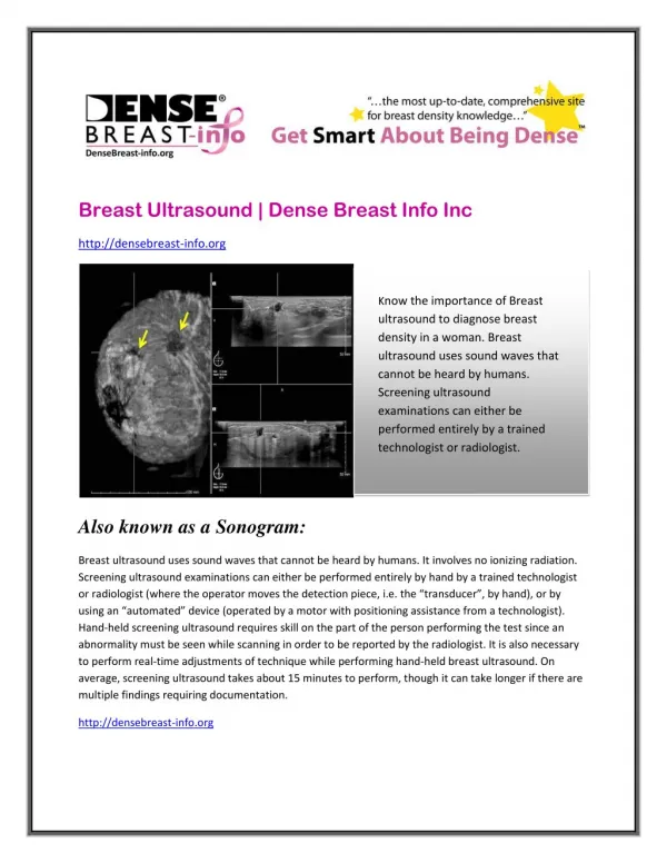
Breast cancer - screening • Self breast examination • Physical examination • Breast ultrasound • Mammography: Mortality can be reduced up to 40% in women, the benefit is greatest in women aged over 50y/o
Breast cancer - screening • Age range: Reduction in mortality is greatest in women aged 50-70% • Frequency of screening: propably between 18 months and 2 years
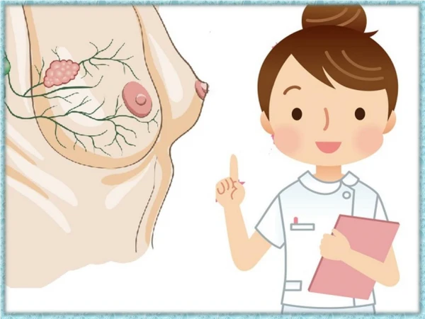
Visual examination in front of a mirror Examination while standing in the shower Examination while lying down Breast Self-examination (BSE)
What are you looking for in BSE • Lumps that are hard, irregular in shape, not painful, and not mobile (in breast or axilla) • A change on the skin of breast • Assymetricity of Bil. breasts or a change of shape of breast • Nipple discharge • Eczematoid change over areola or nipple