
Cells
E N D
Presentation Transcript
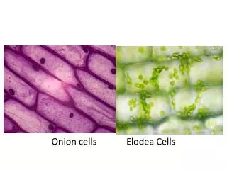
Cells • Cells are small. • There are 100 trillion cells in the body. • They range in size from 7.5 µm = micrometers (micrometer is 1 millionth of a meter) to 250 µm, which is visible to the naked eye. • There are thousands of types of cells, each is specialized for a task: skin, liver, kidney, etc. • Each cell has specialized structures for their function.
Cells Every cell has three things in common: • Metabolic functions (using nutrients such as sugars and oxygen, and creating waste products) • Responds to its environment • Capable of maintaining homeostasis within itself and within the body. HOMEOSTASIS is maintaining a constant and appropriate internal environment, such as temperature, pH, and glucose levels.
Introduction to Cells • All cells have several main components • Plasma membrane • Cytoplasm and cytosol • Nucleus • Organelles (are surrounded by a membrane) • Ribosomes (are not surrounded by a membrane)
Structure of a Generalized Cell Figure 2.1
Cytoplasm and Cytosol CYTOPLASM: the watery liquid inside and outside the organelles, but outside the nucleus. • NEUCLEOPLASM: the liquid inside the nucleus. • CYTOSOL: another liquid that is thicker than water, and is NOT inside the organelles. It is only found outside of the organelles and nucleus. • Cytosol contains the following: • Mostly water • Things dissolved in water (amino acids, sugars like glucose, nucleic acids, and ATP, which is a molecule used for energy). • Cytoskeleton: made up of long protein fibers, extend throughout cytosol. Function of cytoskeleton: 1) Maintains cell shape 2) Movement (such as muscle cell contraction, organelles within the cell, or the cell itself moving around).
Cell Membrane • The cell membrane is semi-permeable to allow only certain things into and out of the cell. • Functions of the Plasma Membrane: • Movement of materials into and out of cell, and acts as a barrier to the external environment • Acts as a site for receiving signals from the rest of the body • Helps hold the cell in place
Plasma (cell) Membrane • The plasma (cell) membrane is made up of two layers of molecules = PHOSPHOLIPIDS. • It’s therefore called a phospholipid bilayer • Phospholipids are amphipathic molecules. • That means they have one end that has an affinity for something and another end that does not have an affinity to that substance. In this case, the affinity is to water. • A substance that likes water is called HYDROPHILIC (likes water). • A region of a molecule that is hydrophilic is called a POLAR region.
Plasma (cell) Membrane • A substance that dislikes water is called HYDROPHOBIC (afraid of water). • A region of a molecule that is hydrophobic is called a NON-POLAR region. • Therefore, the phospholipids, being amphipathic, will have a polar region and a non-polar region. • The polar region is the PHOSPHATE HEADS • The non-polar region is the FATTY ACID TAILS .
Phosphate heads Fatty Acid tails The cell membrane is like a film of oil on water. Is oil flexible? (yes) Is oil strong? (no) But it prevents materials from going across into the water.
Plasma (cell) Membrane • The plasma membrane has proteins in it that are made in the RIBOSOMES and transported to the cell membrane in this case (other proteins are carried elsewhere). • Ribosomes carry out the three functions of the plasma membrane. • Around each organelle is a membrane identical to the plasma membrane except for the proteins. • Each cell has hundreds of membranes. • Ribosomes are not organelles because they do not have a plasma membrane.
The Cell Membrane Phospho-lipid Bilayer Figure 2.2a
Endoplasmic Reticulum • The ER is a network of channels. • Two types: • Rough ER: contains ribosomes • Function of ribosomes is to make proteins. • Smooth ER: no ribosomes • Function is to detoxify chemicals that enter the cell.
ROUGH ENDOPLASMIC RETICULUM (endoplasmic = within cytoplasm; reticulum = network; rough = surface of membrane covered with ribosomes. This is an organelle, but the ribosomes are not. • Function of RER is the synthesis (making) of proteins: a. Membrane proteins b. Proteins for export (such as digestive system enzymes) c. Proteins for use within the cell
SMOOTH ENDOPLASMIC RETICULUM (no ribosomes) Function of SER a. SER is continuous with the rough ER, but lacks ribosomes and has several functions 1) Detoxifies harmful substances (alcohol, drugs, medicines) NOTE: in CSI, when they suspect poisoning, they first look at the SER in the liver. 2) Stores calcium 3) Involved in lipid production (lipid bodies)
The Endoplasmic Reticulum and Ribosomes Figure 2.5
Golgi Complex • When the proteins have finished their journey in the RER, their edges are exposed, and are vulnerable to oxidative damage. Therefore, they first go to the Golgi complex, which puts chemical bonds on the ends of the proteins. • Thus, in the Golgi complex, the proteins are modified and prepared for transport out of the cell. • The Golgi complex is like a Fed-Ex center that packages and ships the proteins that were made in the ribosomes.
Golgi Apparatus Figure 2.8
Vesicles • Vesicles (vacuoles) are bubble-like containers for various substances. Some are created by the end of the Golgi complex: a piece of membrane pinches off, leaving a protein in the vesicle, which carries the protein to the cell membrane, where it merges with the cell membrane, pops, and releases its contents outside of the cell. • Other vesicles are storage containers for food or enzymes.
Vesicles VESICLES: a sphere of membrane with something in it. This is an organelle. Many types: • LYSOSOMES: are sacs of powerful digestive enzymes to dissolve an old organelle, bacteria, or foreign debris. They are also used to commit cell suicide (APOPTOSIS is the term for programmed cell death). • When bacteria enter a cell, the lysosome will fuse with the bacteria and release its enzymes on them to destroy them. • TRANSPORT VESICLES: when material needs to move from RER to Golgi complex, or from Golgi complex to cell membrane, etc. • STORAGE VESICLES:one vesicle may store carbohydrates, one may store lipids, one may store enzymes.
Disorder of Lysosomes • Tay–Sachs disease • A genetic disorder that causes deterioration of mental and physical abilities that commences around six months of age and usually results in death by the age of four. • Caused by insufficient activity of an enzyme needed by lysosomes to break down phospholipids. • The lipids accumulate in the brain.
Mitochondria • Mitochondria are considered the smallest living units in the body because they can make their own energy (ATP). Cells have hundreds of mitochondria. • Function of mitochondria is to make most of the cell’s ATP, which is cellular energy (ATP is an energy source). • Some ATP is made in the cytosol, but most is made in the mitochondria. • NOTE: Mitochondria must have OXYGEN to convert nutrients to ATP for energy.
Mitochondria • Mitochondria – generate most of the cell’s energy (ATP); most complex organelle. • Contains curves known as cristae that can be seen under a microscope. Figure 2.9
Fun Facts Mitochondrial DNA (mtDNA) • Nuclear and mitochondrial DNA are thought to be of separate evolutionary origin, with the mtDNA being derived from the DNA of the bacteria that were engulfed by the early ancestors of today's eukaryotic cells. • mtDNA is inherited from the mother (maternally inherited). • This enables researchers to trace maternal lineage far back in time.
Mitochondrial DNA • Biologists can determine and then compare mtDNA sequences among different species and use the comparisons to build an evolutionary tree for the species examined. • Studies have used mtDNA to trace the ancestry of domestic dogs to wolves. • However, they have recently found that the Sabre-tooth tiger is not the ancestor of the domestic cat.
Nucleus • NUCLEUS: Usually the largest structure in a cell. It does not contain cytoplasm; it is called nucleoplasm. • The nuclear membrane contains pores, called nuclear pores. These allow certain materials into and out of the nucleus. • Functions of the nucleus: • Stores DNA (chromosomes are made up of DNA) • Makes RNA (RNA is the code for making a protein. It is copied from DNA).
Nucleolus • Within a nucleus there are areas that are darker. These are regions of condensed RNA. Remember, the function of the RNA is to carry copies of the genes for proteins to the ribosomes. • The nucleolus is NOT an organelle, but the nucleus is. Don’t get “nucleolus” mixed up with the word “nucleus” on the test. The nucleolus does not contain the DNA; the nucleus does. The nucleolus is within the nucleus, but it does NOT contain DNA. • The nucleolus contains RNA, which is important for protein synthesis. • Do not get nucleus and nucleolus mixed up!
Centrioles • Centrioles are filaments within the cell that function during mitosis. • When the cell goes from metaphase to anaphase of mitosis, the chromatids separate and follow the spindles of the centrioles towards the opposite ends of the cell.
Centrioles Centrioles
Flagellum • Some cells have a flagellum, which is a whip-like tail used to help them move (locomotion). • An example is a sperm cell.
Microvilli • Some cells have microvilli on their cell membrane, which increase the surface area of cells by approximately 600 fold, thus facilitating absorption and secretion.
Cilia • Some cells have cilia, which are small, hair-like structures that can wave back and forth, causing substances to move along across the top of the cell. • For example, the cells of the lungs are lined with cilia, which move mucous up from the lungs so it can be coughed up and swallowed.
Cell Cycle • CELL CYCLE: the life cycle of a cell. Some cells never divide (neurons). • When getting ready to divide, cells undergo MITOSIS to make one cell into two. • Some cells divide rapidly (every few days), some rarely (every 1-2 months), some never.
Stem Cells • STEM CELLS: A population of cells are always available to replace the cells that died. • Muscle stem cells give rise to new muscle cells. • Bone marrow stem cells give rise to new blood cells. • Embryonic stem cells give rise to any type of cells, including neurons (adults don’t have neural stem cells) and pancreatic cells (diabetics don’t have pancreatic stem cells). • Stem cells are named by type + suffix: BLAST • Erythrocyte = RBC. Erythroblast = stem cell that gives rise to an erythrocyte.
Human Cell Division • All cells in our body divide by duplicating their chromosomes and then splitting into two cells, a process called mitosis • Mitosis produces two daughter cells with the same number and kind of chromosomes as the parent cell. • If a parent cell has 46 chromosomes prior to mitosis, how many chromosomes will the daughter cells have? • Answer = 46. • This condition is called diploid (2n).
Sex Cells (Gametes; egg and sperm cells) • After mitosis, sex cells undergo another cell division without duplicating the chromosomes. This is called meiosis: each daughter cell has only half of the chromosomes. • In males, it produces the cells that become sperm • In females, it produces the cells that become eggs. • The sperm and the egg are the sex cells, or gametes. • GAMETES contain half the number of chromosomes compared to the rest of the body cells (23 chromosomes). • This condition is called haploid (n).
Mitosis Stages • Interphase: Chromosomes duplicate stage) • Prophase: Chromosomes shorten and thicken. • Metaphase: Chromosomes line up in the middle of the cell • Anaphase: Chromosomes pull apart • Telophase: Cytoplasm divides in two, forming two daughter cells
MEIOSIS • Meiosis only occurs in the testes and ovaries when they are ready to make an egg cell or a sperm cell. • First, mitosis occurs as normal. • But right after that, the two daughter cells divide again (meiosis), but this time there is no reproduction of the chromosomes.
Crossing Over • During meiosis, when the second cell division is at the metaphase stage, the chromosomes touch each other and exchange a few genes. • The exchange of genetic material between chromatids is calledcrossing-over. • That is what allows for genetic variation.
MEIOSIS • Meiosis results in four daughter cells, each having half the number of chromosomes as the parent cell. • The daughter cells are not genetically identical, and neither is identical to the parent cell. • For example, in MEIOSIS, if the parent cell has 46 chromosomes, the GAMETE will have 23. • It will be haploid (n).
Gametes to Zygote • When a sperm and egg (gametes) combine and contribute their chromosomes, the fertilized egg (called a zygote) will now have 46 chromosomes again. • It will be diploid (2n).
Nondisjunction • Chromosomes can become abnormal if the sister chromosomes do not separate properly during meiosis. This is called nondisjunction.