Chapter 6 Adipose Tissue
Chapter 6 Adipose Tissue. A specialized type of connective tissue in which adipocytes or fat cells predominate. Located in many areas throughout the body, A dipose tissue represents 15–20% of the body weight in men. in women of normal weight, 20–25% of body weight . .


Chapter 6 Adipose Tissue
E N D
Presentation Transcript
A specialized type of connective tissue in which adipocytesor fat cells predominate.Located in many areas throughout the body, Adipose tissue represents 15–20% of the body weight in men.in women of normal weight, 20–25% of body weight.
key regulators of the body’s energy metabolism. Because of a growing worldwide epidemic of obesity and its associated problems, including diabetes and heart disease, adipocytes are now the most widely studied cell of connective tissue.
Adipose tissue is the largest repository of energy (in the form of triglycerides, the neutral fats) in thebody. The other organs that store energy, notably the liver and skeletal muscle, do so in the form of glycogen.However the supply of glycogen is limited and a large store of calories must be mobilized between meals. Because triglycerides are of lower density than glycogen and have a higher caloric value (9.3 kcal/g for triglycerides versus 4.1 kcal/g for carbohydrates), adipose tissue has evolved as a very efficient storage tissue.
Adipocytesthemselves release hormones and a number of important factors. With its unique physical properties, adipose tissue or fat is a poor heat conductor. and it contributes to the thermal insulation of the body. It fills up spaces between other tissues and helps to keep some organs in place. Subcutaneous layers of adipose tissue help to shape the surface of the body, whereas deposits in the form of pads act as shock absorbers, chiefly in the soles and palms.
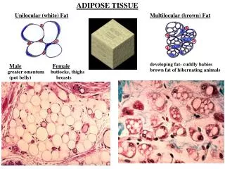
Two types of adipose tissue • with different locations, structures, colors, and pathologic characteristics. • White adipose tissue, the more common type, is composed of cells that, when completely developed, contain one large central droplet of whitish-yellow fat in their cytoplasm. • Brown adipose tissue contains cells with multiple lipid droplets interspersed among abundant mitochondria, which give these cells the darker appearance. Both types of adipose tissue have a rich blood supply.
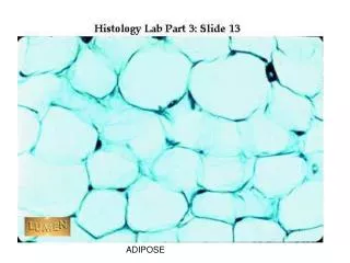
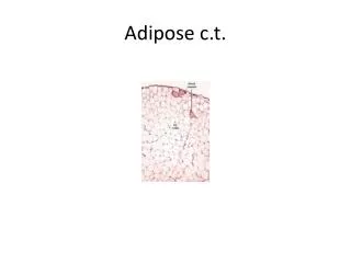
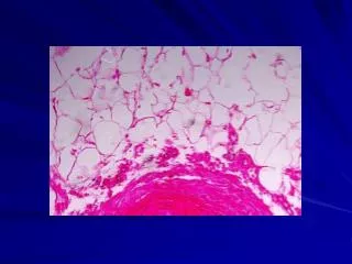
White Adipose Tissue • Specialized for long-term energy storage, white adipose cells are spherical when isolated but are polyhedral when closely packed in adipose tissue. Each cell is very large, between 50 and 150 m in diameter and contains one huge droplet of lipid that makes up 85% of the cell's weight. • called unilocular because triglycerides are stored in a single locus. Since lipid is removed from cells by the alcohol and xylene used in routine histological techniques,
A unilocularadipocyte appears in standard microscope preparations as a thin ring of cytoplasm surrounding the empty vacuole left by the dissolved lipid droplet, sometimes referred to as the signet ring cell.The large droplet causes these cells to have eccentric and flattened nuclei.The rim of cytoplasm that remains after removal of the stored triglycerides may rupture and collapse, distorting the tissue structure.
The thickest portion of the cytoplasm surrounds the nucleus of these cells and contains a Golgi apparatus, mitochondria, poorly developed cisternae of the rough ER, and free polyribosomes. • The rim of cytoplasm surrounding the lipid droplet contains cisternae of smooth ER and numerous pinocytotic vesicles. • TEM studies reveal that each adipose cell usually possesses minute lipid droplets in addition to the single large droplet seen with the light microscope; the droplets are not enveloped by a membrane but show many vimentin intermediate filaments in their periphery. • Each adipose cell is surrounded by a thin external or basal lamina.
White adipose tissue is subdivided into incomplete lobules by a partition of connective tissue containing a rich vascular bed and nerve network. Fibroblasts, macrophages, and other cells make up about half the total number of cells. Reticular fibers form a fine interwoven network that supports individual fat cells and binds them together. Although blood vessels are not always apparent in tissue sections, adipose tissue is richly vascularized.
The color of freshly dissected white adipose tissue depends on the diet and varies from white to bright yellow, due mainly to the presence of carotenoids dissolved in the fat droplets. Almost all adipose tissue in adults is of this type and it is found in many organs throughout the body. Age and gender determine the distribution and density of adipose deposits.
In the newborn, white adipose tissue has a more uniform thickness throughout the body. As the child matures, the tissue tends to disappear from some parts of the body and increase in others. Its distribution is partly regulated by sex hormones, which control adipose deposition in the breasts and thighs.
Storage & Mobilization of Lipids • The white adipose tissue is a large depot of energy for the organism. • The lipids stored in adipose cells are chiefly triglycerides, ie, esters of fatty acids and glycerol. • Triglycerides stored by these cells originate in dietary fats brought to adipocytes as circulating chylomicrons, • in triglycerides synthesized in the liver and transported to adipose tissue in the form of very low-density lipoproteins (VLDLs), and by the local synthesis of free fatty acids and glycerol from glucose to form triglycerides.
Chylomicrons (=small) are particles up to 3 m in diameter, formed in intestinal epithelial cells and transported in blood plasma and mesenteric lymph. They consist of a central core, composed mainly of triglycerides and a small quantity of cholesterol esters, surrounded by a stabilizing monolayer consisting of apolipoproteins, cholesterol, and phospholipids. VLDL are smaller than chylomicrons (providing a greater surface-to-volume ratio) and have proportionately more lipid in their surface layer. VLDL also have different apolipoproteins at the surface and contain a higher proportion of cholesterol esters to triglycerides than do chylomicrons.
Lipid storage and mobilization from adipocytes. Triglyceridesare transported by lymph and blood from the Intestine and liver in lipoprotein complexes known as chylomicrons(Chylo) and very low—density lipoproteins (VLDLs). In capillary endothelial cells of adipose tissue, these complexes are partly broken down by lipoprotein lipase, releasing free fatty acids and glycerol.
The free fatty acids diffuse from the capillary into the adipocyte, where they are re—esterified to glycerol phosphate, forming triglycerides. These resulting triglycerides are stored in droplets until needed. Norepinephrinefrom nerve endings stimulates the cyclic AMP (cAMP) system, which activates hormone—sensitive lipase to hydrolyze the stored triglycerides to free fatty acids and glycerol. These substances diffuse into the capillary, where free fatty acids bind to albumin for transport to distant sites for use as an energy source.
Adipose tissue also functions as an important endocrine organ. Adipocytes are the sole source of the 16 kDa polypeptide hormone leptin (Gr. leptos, thin), a "satiety factor" with target cells in the hypothalamus and other organs, which regulates the appetite under normal conditions and participates in regulating the amount of adipose tissue.
Although all white adipose tissue appears histologically and physiologically similar, differences in gene expression have been noted between • visceral deposits (in the abdomen) • and subcutaneous deposits of white fat. • Such differences may be important in the medical risks of obesity; it is well-established that increased visceral adipose tissue raises the risk of diabetes and cardiovascular disease whereas increased subcutaneous fat does not. • The release of visceral fat products directly to the portal circulation and liver may also influence the medical importance of this form of obesity.
In response to body needs, lipids are mobilized uniformly in all parts of the body, although adipose tissue in the palms, soles, and retroorbital fat pads resists long periods of starvation. After periods of starvation, most unilocularadipocytes lose nearly all their fat and become polyhedral or spindle-shaped cells with very few lipid droplets.
Histogenesis of White Adipose Tissue • Like the fiber-producing cells of connective tissue, adipocytes undergo differentiation from embryonic mesenchymal cells. • Such differentiation is first seen with the appearance of lipoblasts. • Early lipoblasts have the appearance of fibroblasts but are able to accumulate fat in their cytoplasm. • Lipid accumulations are isolated from one another at first but soon fuse to form the single larger droplet that is characteristic of unilocular adipose tissue cells.
Brown Adipose Tissue The color of brown adipose tissue or brown fat is due to both the numerous mitochondria (containing coloredcytochromes) scattered through the adipocytes and the large number of blood capillaries in this tissue. Adipocytesof brown fat contain many small lipid inclusions and are therefore called multilocular. The many small lipid droplets, abundant mitochondria, and rich vasculature all help mediate this tissue's principal function of heat production. In comparison with white adipose tissue, which is present throughout the body, brown adipose tissue has a much more limited distribution.
Cells of brown adipose tissue cells are polygonal and generally smaller than cells of white adipose tissue but their cytoplasm contains a great number of lipid droplets of various sizes. These adipocytes have spherical and central nuclei and the numerous mitochondria have abundant long cristae.
Brown adipose tissue resembles an endocrine gland in that its cells assume an almost epithelial arrangement closely associated with blood capillaries. • The tissue is subdivided by partitions of connective tissue into lobules that are better delineated than the lobules of white adipose tissue. • Cells of this tissue receive direct sympathetic innervation.
Function of Brown Adipocytes • The main function of the multilocular adipose cells is to produce heat by nonshiveringthermogenesis. • The physiology of multilocular adipose tissue is best understood in the study of hibernating species. • In animals ending their hibernation period, or in newborn mammals (including humans) that are exposed to an environment colder than the mother's uterus, nerve impulses liberate norepinephrine into brown adipose tissue.
unlike white fat, liberated fatty acids of multilocularadipocytes are quickly metabolized, with a consequent increase in oxygen consumption and heat production, elevating the temperature of the tissue and warming the blood passing through it. Heat production is increased in these cells because the mitochondria have in their inner membrane a transmembrane protein called thermogeninor uncoupling protein (UCP-1), a marker unique to brown fat.
Thermogenin permits the backflow of protons previously transported to the intermembranous space without passing through the ATP-synthetase system in the mitochondrial globular units. • Consequently, the energy generated by proton flow is not used to synthesize ATP and is dissipated as heat. Warmed blood circulates throughout the body, distributing the heat and carrying fatty acids not metabolized in the adipose tissue for use elsewhere.
Histogenesis of Brown Adipose Tissue • Brown adipose tissue also develops from embryonic mesenchyme, that emerges earlier than white fat during fetal development. • In humans the amount of brown fat is maximal relative to body weight at birth, when nonshiveringthermogenesis is most needed. • The tissue largely disappears (by involution) or is replaced by white fat during childhood. • In adults it is found only in scattered areas, especially around the kidneys and adrenal glands, the aorta, and mediastinum.
The number of brown adipocytes increases again during cold adaptation, Besides stimulating thermogenic activity, autonomic nerves also promote brown adipocyte differentiation and prevent apoptosis in mature brown cells.