Download
1 / 30
310 likes | 801 Views
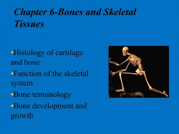
Chapter 6 Bones and Skeletal Tissue. Angela Peterson-Ford, PhD apetersonford@collin.edu. 6. Bones and Skeletal Tissues Part 1. Skeletal Cartilage. Contains no blood vessels or nerves Surrounded by the perichondrium (dense irregular connective tissue) that resists outward expansion

E N D
Chapter 6Bones and Skeletal Tissue Angela Peterson-Ford, PhD apetersonford@collin.edu
6 Bones and Skeletal Tissues Part 1
Skeletal Cartilage • Contains no blood vessels or nerves • Surrounded by the perichondrium (dense irregular connective tissue) that resists outward expansion • Three types – hyaline, elastic, and fibrocartilage
Hyaline Cartilage • Provides support, flexibility, and resilience • Is the most abundant skeletal cartilage • Is present in these cartilages: • Articular – covers the ends of long bones • Costal – connects the ribs to the sternum • Respiratory – makes up the larynx and reinforces air passages • Nasal – supports the nose
Elastic Cartilage • Similar to hyaline cartilage but contains elastic fibers • Found in the external ear and the epiglottis
Fibrocartilage • Highly compressed with great tensile strength • Contains collagen fibers • Found in menisci of the knee and in intervertebral discs
Growth of Cartilage • Appositional – cells in the perichondrium secrete matrix against the external face of existing cartilage • Interstitial – lacunae-bound chondrocytes inside the cartilage divide and secrete new matrix, expanding the cartilage from within • Calcification of cartilage occurs • During normal bone growth • During old age
Bones and Cartilages of the Human Body Figure 6.1
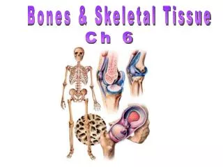
Classification of Bones • Axial skeleton – bones of the skull, vertebral column, and rib cage • Appendicular skeleton – bones of the upper and lower limbs, shoulder, and hip
Classification of Bones: By Shape • Long bones – longer than they are wide (e.g., humerus) Figure 6.2a
Classification of Bones: By Shape • Short bones • Cube-shaped bones of the wrist and ankle • Bones that form within tendons (e.g., patella) Figure 6.2b
Classification of Bones: By Shape • Flat bones – thin, flattened, and a bit curved (e.g., sternum, and most skull bones) Figure 6.2c
Classification of Bones: By Shape • Irregular bones – bones with complicated shapes (e.g., vertebrae and hip bones) Figure 6.2d
Function of Bones • Support – form the framework that supports the body and cradles soft organs • Protection – provide a protective case for the brain, spinal cord, and vital organs • Movement – provide levers for muscles • Mineral storage – reservoir for minerals, especially calcium and phosphorus • Blood cell formation – hematopoiesis occurs within the marrow cavities of bones
Bone Markings • Bulges, depressions, and holes that serve as: • Sites of attachment for muscles, ligaments, and tendons • Joint surfaces • Conduits for blood vessels and nerves
Bone Markings: Projections – Sites of Muscle and Ligament Attachment • Tuberosity – rounded projection • Crest – narrow, prominent ridge of bone • Trochanter – large, blunt, irregular surface • Line – narrow ridge of bone
Bone Markings: Projections – Sites of Muscle and Ligament Attachment • Tubercle – small rounded projection • Epicondyle – raised area above a condyle • Spine – sharp, slender projection • Process – any bony prominence
Bone Markings: Projections – Projections That Help to Form Joints • Head – bony expansion carried on a narrow neck • Facet – smooth, nearly flat articular surface • Condyle – rounded articular projection • Ramus – armlike bar of bone
Bone Markings: Depressions and Openings • Meatus – canal-like passageway • Sinus – cavity within a bone • Fossa – shallow, basinlike depression • Groove – furrow • Fissure – narrow, slitlike opening • Foramen – round or oval opening through a bone
Gross Anatomy of Bones: Bone Textures • Compact bone – dense outer layer • Spongy bone – honeycomb of trabeculae filled with yellow bone marrow
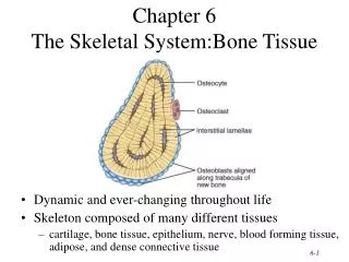
Structure of Long Bone • Long bones consist of a diaphysis and an epiphysis • Diaphysis • Tubular shaft that forms the axis of long bones • Composed of compact bone that surrounds the medullary cavity • Yellow bone marrow (fat) is contained in the medullary cavity
Structure of Long Bone • Epiphyses • Expanded ends of long bones • Exterior is compact bone, and the interior is spongy bone • Joint surface is covered with articular (hyaline) cartilage • Epiphyseal line separates the diaphysis from the epiphyses
Structure of Long Bone Figure 6.3
Bone Membranes • Periosteum – double-layered protective membrane • Outer fibrous layer is dense regular connective tissue • Inner osteogenic layer is composed of osteoblasts and osteoclasts • Richly supplied with nerve fibers, blood, and lymphatic vessels, which enter the bone via nutrient foramina • Secured to underlying bone by Sharpey’s fibers • Endosteum – delicate membrane covering internal surfaces of bone
Structure of Short, Irregular, and Flat Bones • Thin plates of periosteum-covered compact bone on the outside with endosteum-covered spongy bone (diploë) on the inside • Have no diaphysis or epiphyses • Contain bone marrow between the trabeculae
Structure of a Flat Bone Figure 6.4
Location of Hematopoietic Tissue (Red Marrow) • In infants • Found in the medullary cavity and all areas of spongy bone • In adults • Found in the diploë of flat bones, and the head of the femur and humerus
Microscopic Structure of Bone: Compact Bone • Haversian system, or osteon – the structural unit of compact bone • Lamella – weight-bearing, column-like matrix tubes composed mainly of collagen • Haversian, or central canal – central channel containing blood vessels and nerves • Volkmann’s canals – channels lying at right angles to the central canal, connecting blood and nerve supply of the periosteum to that of the Haversian canal
Microscopic Structure of Bone: Compact Bone • Osteocytes – mature bone cells • Lacunae – small cavities in bone that contain osteocytes • Canaliculi – hairlike canals that connect lacunae to each other and the central canal
Microscopic Structure of Bone: Compact Bone Figure 6.6a, b