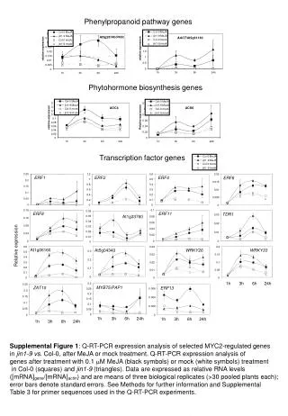
Download

1 / 37
370 likes | 527 Views
Molecular Regulation of Development. Growth factor signaling, Hox genes and the body plan. 张咸宁 zhangxianning@zju.edu.cn Tel: 13105819271; 88208367 Office: A705, Research Building 2012/09. 出生缺陷( Birth defect ):即先天性疾病.

E N D
Molecular Regulation of Development. Growth factor signaling,Hox genes and the body plan 张咸宁 zhangxianning@zju.edu.cn Tel:13105819271; 88208367 Office: A705, Research Building 2012/09
出生缺陷(Birth defect):即先天性疾病 • Malformation is a primary morphologic defect of an organ or body part resulting from an intrinsically abnormal developmental process (e.g., cleft lip, polydactyly). • Dysplasia is a primary defect involving abnormal organization of cells into tissue (e.g., vascular malformation). • Sequence is a primary defect with its secondary structural changes (e.g., Pierre Robin sequence, a disorder in which a primary defect in mandibular development produces a small jaw, secondary glossoptosis, and a cleft palate) • Syndrome is a pattern of multiple primary malformations with a single etiology (e.g., trisomy 13 syndrome). • Deformation is alteration of the form, shape, or position of a normally formed body part by mechanical forces. It usually occurs in the fetal period, not in embryogenesis. It is a secondary alteration. It can be extrinsic, as in oligohydramnios (reduced amniotic fluid), or intrinsic, as in congenital myotonic dystrophy. • Disruption is a morphological defect of an organ, part of an organ, or a larger region of the body resulting from the extrinsic breakdown of, or interference with, an originally normal developmental process. It is a secondary malformation (e.g., secondary limb defect resulting from a vascular event).
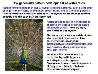
Three questions: 1. How is dorsal-ventral (D-V背腹) cell differentiation regulated by morphogen(形态发生素)gradients(梯度). BMP(骨形态发生蛋白) signal transduction. 2. Hox is the the antero-posterior (A-P前后) pattern regulated by Hox genes? - Colinearity(共线性) - Activation by retinoic acid(视黄酸). Retinoid receptors. 3. How have conserved(保守)gene networks affected Evolution and Development? Evo-Devo.
1. During development groups of inducing cells called organizing centers secrete graded growth factor signals. The concentration gradient of a “morphogen” can induce multiple cell differentiation choices. Fig. 1
The best example of a morphogen is the gradient of BMP signaling that controls D-V tissue differentiation. Fig. 3a
Mesoderm(中胚层) cell differentiation is determined by a morphogen gradient of BMP. BMP signaling ventral dorsal Lateral plate Somite Notochord BMPs are members of the TGFβ superfamily. Fig. 3b
Genes specifically expressed in the dorsal blastopore lip (胚孔唇Spemann organizer) of the gastrula(原肠胚)were cloned. Organizer-specific Genes Fig. 4a
Chordin(脊索发生素)mRNA is expressed in Spemann’s organizer. Chordin protein is secreted and diffuses in the embryo. Fig. 4b
Chordin is an antagonist(拮抗剂)that binds BMP growth factors in the extracellular space, inhibiting binding to cell surface receptors. Chordin establishes a BMP4 activity gradient at gastrula. Another protein, Noggin(头发生素), has similar activity. Secreted antagonists diffuse and are used in development to generate morphogen gradients. Chordin inhibits Fig. 5
Signal transduction: membrane receptors transduce the signal so that transcription factors are activated through phosphorylation. TGFβ family members (30 different ligands in humans) activate cell surface receptors (Serine-Threonine kinases). Xnr BMP4 Fig. 6a
Visualizing a morphogen gradient: phosphorylated Smad1 forms a gradient, maximal in the ventral, in the Xenopus(爪蟾)gastrula(原肠胚) Dorsal Ventral Blastopore Transverse section at the level of white arrows Side view Fig. 7a
Epidermis CNS Dorsal Ventral Spemann’s Organizer Mesoderm Endoderm At gastrula a gradient of BMP4 is established by a ventral source of BMP4 and a dorsal source of Chordin and Noggin, two BMP antagonists secreted by the dorsal organizing center. Fig. 7b
The BMP gradient induces different tissues in mesoderm中胚层 and ectoderm外胚层 (because the DNA-binding partners are different) Ectoderm differentiation Mesoderm differentiation BMP signaling BMP signaling Neural crest Lateral plate Somite Notochord Epidermis CNS Fig. 7c
Conclusion:a morphogen gradient can be generated by a source of growth factor (such as BMP) or by a localized source of inhibitor (such as Chordin). Both mechanisms are used. This is how organizing centers work in embryonic induction. Fig. 8
Cell-cell communication is controlled by surprisingly few signal transduction pathways: TGFβ/BMP Serine/Threonine kinase receptors Receptor Tyrosine kinases such as FGF, EGF, IGF, Insulin Wnts Sonic Hedgehog Notch G protein-coupled receptors (7-transmembrane receptors) Nuclear hormone receptors Only a few signaling pathways pattern the embryo, but there are hundreds of differentiated cell types in the human body. The same signals can trigger different types of cell differentiation responses in cells of different developmental history (because of different DNA-binding partners). Fig. 9
A. Hox genes: colinearity between the gene order in genomic DNA and the body plan B. Hox genes and Retinoic acid C. Hox genes in Evolution and Development (Evo-Devo) A-P patterning outline: Fig. 10
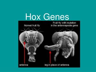
2. Hox genes and the development of body plans Homeotic transformations(同源异型转化) in humans. A cervical vertebra(颈椎)transformed into a thoracic one with ribs(胸肋). Fig. 11
Edward B. Lewis Homeotic genes specify body segment identity in Drosophila. Lewis predicted Hox genes would be duplicated. Fig. 13
Homeobox(同源〔异型〕框)refers to nucleic acid. Homeodomain(同源〔异型〕域)refers to protein. The homeodomain is a 60 aa helix-turn-helix DNA-binding domain that is very conserved during evolution. It fits into the major groove of the DNA. Define Hox, homeobox The term homeobox is reserved for the nucleic acid sequences that encode homeodomains. Since they are highly conserved, and can be detected by low-stringency hybridization across species. Fig. 14
Hox complexes are conserved between Drosophila and mammals (from de Robertis et al., Scientific American, 1990) Fig. 15
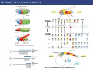
Vertebrates have 4 Hox complexes, with about 10 genes each. They display colinearity(共线性): a) Spatial(空间)colinearity: the more anteriorly(前部)expressed genes are in one end, the more posterior ones at the other end of the gene complex. b) Temporal(时间)colinearity: genes on one end of the complex are expressed first, those on the other (posterior。后部) end are turned on last. c) Anterior Hox genes are activated sequentially by retinoic acid. Hox genes can be aligned in 13 groups of paralogues (种内同源基因。that were duplicated twice). Fig. 16
Extensive conservations between Drosophila and the four human Hox complexes High RA response Low RA response From de Robertis E.M. Evo-Devo: Variations on Ancestral themes. Cell 132, 185-195 (2008) Fig. 17
Hox-C6 protein is seen in 8 thoracic segments of the mouse embryo. Translation is blocked in the tail region, probably through the action of microRNAs. The inset shows that Hox-C6 mRNA is expressed all the way to the tip of the tail. From De Robertis, Cell 132, 185-195 (2008) Fig. 18
Spatial and temporal colinearity: order of Hox genes in DNA follows the antero-posterior body axis. Why have Hox genes stayed together in a complex? Fig. 19
Hox knockouts in mice cause homeotic transformations, in this case an extra rib in the lumbar region(HoxC-8 mutant). Treatment with retinoic acid can also cause lumbar ribs(腰肋). Your patient this week has 13 ribs. Fig. 20
B. Retinoic acid activates HOX genes sequentially in cultured human teratocarcinoma(畸胎癌)cells mRNA amount Fig. 21
How do Retinoic acid receptors work? LBD DBD Dimerization ligand coactivator Fig. 22
Retinoic acid receptor is a DNA-binding protein that works as a ligand-activated transcription factor. Many hydrophobic hormone receptors work in this way. Nuclear receptors work very differently from cell surface receptors. (RA) RA Fig. 23
Hox complexes have a retinoic acid receptor response element (RARE) in the DNA before paralogue 1. This DNA enhancer element controls expression of many genes in the complex. In retinoic acid teratogenesis, Hox gene expression borders move into more anterior regions. RARE Fig. 24
Pharyngeal arch 1 does not express any Hox gene. It gives rise to maxillary(上颌骨)and mandibular(下颌骨)structures. Retinoic acid can cause cleft palate(腭裂)and micrognathia(下颌) Fig. 25
III. The common ancestor Urbilateria(原生两侧对称动物)used Hox genes and Chordin/BMP to pattern the embryo. 30 of the 35 animal phyla(门)are bilaterans(两侧对称动物). BMP Hox Chd Fig. 26
Evo-Devo: Urbilateria had a Hox gene complex. Developmental control genes placed evolutionary constraints on the types of animal shapes that evolved by Natural Selection(自然选择). High RA response Low RA response From: de Robertis EM, Evo-Devo: Variations on Ancestral themes. Cell 132, 185-195 (2008) Fig. 27
Overview Langman’s Medical Embryology Fig. 28
Acknowledge(PPT特别鸣谢!) • UCLA David Geffen School of Medicine • www.medsch.ucla.edu/ANGEL/ • Prof. RobertisE(UCLA), et al.