Download
1 / 38
390 likes | 535 Views
This presentation delves into the complex world of gene regulation, highlighting how cells respond dynamically to their environment. It covers the fixed nature of the genome contrasted with the dynamic behavior of cells, emphasizing gene expression variation influenced by cell type, cycle, and external conditions. Key processes such as transcription, translation, and the importance of transcription factors and regulatory elements are discussed. Additionally, the presentation explores the differences between eukaryotic and prokaryotic gene structures, including splicing and codon usage.

E N D
Genes: Regulation and Structure Many slides from various sources, including S. Batzoglou,
Cells respond to environment Various external messages Heat Responds to environmental conditions Food Supply
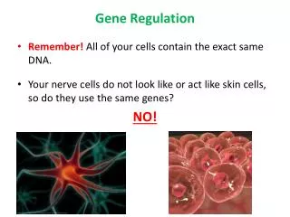
Genome is fixed – Cells are dynamic • A genome is static • Every cell in our body has a copy of same genome • A cell is dynamic • Responds to external conditions • Most cells follow a cell cycle of division • Cells differentiate during development
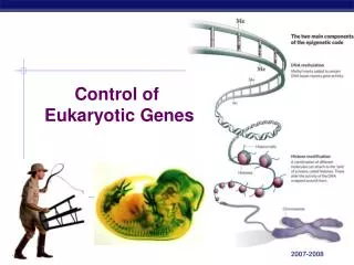
Gene regulation • Gene regulation is responsible for dynamic cell • Gene expression varies according to: • Cell type • Cell cycle • External conditions • Location
Where gene regulation takes place • Opening of chromatin • Transcription • Translation • Protein stability • Protein modifications
Transcriptional Regulation • Strongest regulation happens during transcription • Best place to regulate: No energy wasted making intermediate products • However, slowest response time After a receptor notices a change: • Cascade message to nucleus • Open chromatin & bind transcription factors • Recruit RNA polymerase and transcribe • Splice mRNA and send to cytoplasm • Translate into protein
Transcription Factors Binding to DNA Transcription regulation: Certain transcription factors bind DNA Binding recognizes DNA substrings: Regulatory motifs
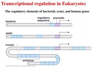
Promoter and Enhancers • Promoter necessary to start transcription • Enhancers can affect transcription from afar
Regulation of Genes Transcription Factor (Protein) RNA polymerase (Protein) DNA Gene Regulatory Element
Regulation of Genes Transcription Factor (Protein) RNA polymerase DNA Regulatory Element Gene
Regulation of Genes New protein RNA polymerase Transcription Factor DNA Regulatory Element Gene
Example: A Human heat shock protein --158 0 HSE CCAAT AP2 AP2 CCAAT SP1 SP1 TATA • TATA box: positioning transcription start • TATA, CCAAT: constitutive transcription • GRE: glucocorticoid response • MRE: metal response • HSE: heat shock element GENE promoter of heat shock hsp70
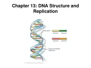
DNA transcription RNA translation Protein Gene expression CCTGAGCCAACTATTGATGAA CCUGAGCCAACUAUUGAUGAA PEPTIDE
Eukaryotes vs Prokaryotes • “Typical” human & bacterial cells drawn to scale. • Eukaryotic cells are characterized by membrane-bound compartments, which are absent in prokaryotes. Brown Fig 2.1 BIOS Scientific Publishers Ltd, 1999
Prokaryotic genes – searching for ORFs. • Small genomes have high gene density Haemophilus influenza – 85% genic • No introns • Operons One transcript, many genes • Open reading frames (ORF) – contiguous set of codons, start with Met-codon, ends with stop codon.
Example of ORFs. There are six possible ORFs in each sequence for both directions of transcription.
Eukaryotes vs Prokaryotes • “Typical” human & bacterial cells drawn to scale. • Eukaryotic cells are characterized by membrane-bound compartments, which are absent in prokaryotes. Brown Fig 2.1 BIOS Scientific Publishers Ltd, 1999
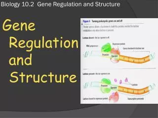
Gene structure intron1 intron2 exon2 exon3 exon1 transcription splicing translation Codon: A triplet of nucleotides that is converted to one amino acid exon = protein-coding intron = non-coding
Gene structure intron1 intron2 exon2 exon3 exon1 transcription splicing translation exon = coding intron = non-coding
Exon 3 Exon 1 Exon 2 Intron 1 Intron 2 5’ 3’ Stop codon TAG/TGA/TAA Start codon ATG Finding genes Splice sites
atg caggtg ggtgag cagatg ggtgag cagttg ggtgag caggcc ggtgag tga
0. We can sequence the mRNA • Expressed Sequence Tag (EST) sequencing is expensive • It has some false positive rates (aberrant splicing) • The method sequences all RNAs and not just those that code for genes • This is difficult for rare genes (those that are expressed rarely or in low quantities. • Still this is an invaluable source of information (when available)
Biology of Splicing (http://genes.mit.edu/chris/)
1. Consensus splice sites Donor: 7.9 bits Acceptor: 9.4 bits (Stephens & Schneider, 1996) (http://www-lmmb.ncifcrf.gov/~toms/sequencelogo.html)
2. Recognize “coding bias” • Each exon can be in one of three frames ag—gattacagattacagattaca—gtaag Frame 0 ag—gattacagattacagattaca—gtaag Frame 1 ag—gattacagattacagattaca—gtaag Frame 2 Frame of next exon depends on how many nucleotides are left over from previous exon • Codons “tag”, “tga”, and “taa” are STOP • No STOP codon appears in-frame, until end of gene • Absence of STOP is called open reading frame (ORF) • Different codons appear with different frequencies—codingbias
2. Recognize “coding bias” Amino Acid SLC DNA codons Isoleucine I ATT, ATC, ATA Leucine L CTT, CTC, CTA, CTG, TTA, TTG Valine V GTT, GTC, GTA, GTG Phenylalanine F TTT, TTC Methionine M ATG Cysteine C TGT, TGC Alanine A GCT, GCC, GCA, GCG Glycine G GGT, GGC, GGA, GGG Proline P CCT, CCC, CCA, CCG Threonine T ACT, ACC, ACA, ACG Serine S TCT, TCC, TCA, TCG, AGT, AGC Tyrosine Y TAT, TAC Tryptophan W TGG Glutamine Q CAA, CAG Asparagine N AAT, AAC Histidine H CAT, CAC Glutamic acid E GAA, GAG Aspartic acid D GAT, GAC Lysine K AAA, AAG Arginine R CGT, CGC, CGA, CGG, AGA, AGG Stop codons Stop TAA, TAG, TGA Can map 61 non-stop codons to frequencies & take log-odds ratios
Approaches to gene finding • Homology • Procrustes • Ab initio • Genscan, Genie, GeneID • Comparative • TBLASTX, Rosetta • Hybrids • GenomeScan, GenieEST, Twinscan, SLAM…
intron exon exon intron intergene exon intergene HMMs for gene finding GTCAGAGTAGCAAAGTAGACACTCCAGTAACGC
T A A T A T G T C C A C G G G T A T T G A G C A T T G T A C A C G G G G T A T T G A G C A T G T A A T G A A Exon1 Exon2 Exon3 GHMM for gene finding duration
Better way to do it: negative binomial • EasyGene: Prokaryotic gene-finder Larsen TS, Krogh A • Negative binomial with n = 3
Splice Site Models • WMM: weight matrix model = PSSM (Staden 1984) • WAM: weight array model = 1st order Markov (Zhang & Marr 1993) • MDD: maximal dependence decomposition (Burge & Karlin 1997) decision-tree like algorithm to take significant pairwise dependencies into account
Donor site 5’ 3’ Position % Splice site detection