Download

1 / 31
310 likes | 327 Views
This paper discusses various segmentation techniques for classifying brain tissues on MR images, including edge-based, region-based, and hybrid techniques. It also explores the challenges and evaluation methods for these techniques.

E N D
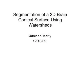
Segmentation of Brain Tissues on MR ImagesUsing Data Driven Techniques Ali Hojjat1, Judit Sovago2, Alan Colchester1 1University of Kent, Canterbury 2Karolinska Institute, Stockholm PVEO Project Meeting Budapest, 11-12 June 2004
Classification of segmentation techniques Segmentation techniques Design based on different principles, eg. Edge based- Use local changes (gradient) – Problem: Very sensitive to noise Region based- Use homogeneity to segment the image • Region growing techniques – Problem of overgrowing • Statistical analysis in feature space like histogram – Problem: lack of spatial information Combined region and edge based techniques Atlas Based- Geometric model of the image – Problem: Relies on the performance of the co-registration step. Hybrid techniques- Combination of two or more of the segmentation techniques.
MR acquisition problems affecting segmentation • Noise • Spatial resolution and partial volume effect • Low resolution • Anisotropic sampling • Intensity inhomogeneity • Motion artefacts
Evaluation of segmentation techniques • Evaluating complete systems in an application domain requires proper definition of application goals and of criteria by which performance is to be judged • Evaluation of algorithms in isolation is difficult • “Gold standard” usually not available • For specific task we might need more than one segmentation algorithm to cover the wide categories of images.
SPM Co-register the image with an atlas using an affine transformation. Perform Cluster Analysis with a modified Mixture Model and a-priori information about the likelihoods of each voxel being one of a number of different tissue types. Do a "cleanup" of the partitions Write the segmented images. The names of these images have "_seg1", "_seg2" & "_seg3" appended to the name of the first image passed.
Pixel Number (in order of linking) Growing process: The grey values of successive points joining the region are shown bellow:
Some definitions: A region containing 20 pixels (shown in black and green). External boundary, EB • External boundary (EB) = regions boundary • Internal boundary (IB) = outermost pixels inside the region • Peripheral contrast = (Mean IB) / (Mean EB)
Grey level (top) and PC (bottom) mapping when region growing started inside the scalp.
Original T1 MRI Flowchart: Segmentation of scalp Seed point In scalp Region growing Find maximum Peripheral contrast Segmented scalp
Grey level (top) and peripheral contrast (bottom) during the growing process for two conditions, with and without the mask.
Sample MR image MR image of an elderly patient with cerebral atrophy. Segmented WM, GM and CSF are shown in Grey, dark grey and white, respectively.
3-D Visualisation of the brain Left Right
BrainSeg Summary • It needs manually selected seed points in the scalp and in the brain • Works well on abnormal as well as normal images • Speed of segmentation of every structure is about 3-4 minutes • Total speed would be about 10 minutes. The speed may vary according to the resolution of the input image and the level of artefacts.
Practical issues related to manual segmentation Questions: Should we use anatomical knowledge to segment a region or only rely on intensity values? What is the best decision when the distance between two parts of a sulcus is very close to zero (touching each other, or less than one pixel)? Crack between pixels might be a good idea, but we should go to subpixel level. Should we rely mainly on our anatomical knowledge when partial volume affects the intensity?
RG BrainSeg SPM
Performance of automatic segmentation technique for brain (WM + GM) tissue BrainSeg
Rate of falsely detected voxels in every slice for the two segmentation techniques BrainSeg
Voxels which are missed by both techniques are shown in green SPM RG BrainSeg
Voxels which are falsely segmented by the two techniques are shown in red SPM RG BrainSeg
Issues related to segmentation of WM WM segmentation is more difficult. Boundary between “WM” and “GM” is not clear. Segmenting tissues changed from their original form,like “WM” lesions, is difficult. Should we segment the “WM” lesions as “WM”, “GM” or separate tissue?
Performance of automatic segmentation technique for WM tissue BrainSeg
Rate of falsely detected voxels in every slice for the two segmentation technique BrainSeg
BrainSeg False positive points (in orange) in the WM overlaid on manual segmentation Original image Manual SPM
BrainSeg –FP & MP SPM -FP & MP False positive (in green) and missed points (in purple) in whole brain Original image Manual
Summary • Manual segmentation of images is very difficult. • Performance of SPM and BrainSeg techniques are very similar. • SPM can segment GM structures (in basal ganglia) better than BrainSeg. • BrainSeg performs better around the cortex. • BrainSeg is three times faster than SPM. • Evaluation should be extended to a larger number of subjects. • We plan to compare the results of PVE correction using different techniques against manual segmentation.