Download

1 / 108
1.09k likes | 1.3k Views
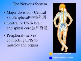
Unit 7– Nervous Tissue & Senses. Central Nervous System (_____). CNS. Brain (100 billion neurons). I. Organization of The Nervous System A. Structural Organization of the Nervous System. Spinal Cord (100 million neurons).

E N D
Central Nervous System (_____) CNS Brain (100 billion neurons) I. Organization of The Nervous System A. Structural Organization of the Nervous System Spinal Cord (100 million neurons)
Peripheral Nervous System (_____) – all nervous tissue outside of CNS PNS 12 Pairs of Cranial Nerves 31 pairs of spinal nerves Ganglia Other nerves
Peripheral Nervous System (PNS) Sensory Receptors in skin & other places
B. Functions of the Nervous System & Types of Neurons that help with Nervous System Functions: 1. _____________________ different types of stimuli from internal and external environment. This is done by: a. ______________________- neurons that carry sensory information from nerves to the CNS or from a lower to a higher level in the CNS Senses Afferent Neurons
Integrates Interneurons 2. _____________________ information by analyzing and storing some of it. This is done by: a. ____________________ - nerve cells whose axons extend for a short distance and contact nearby neurons in the brain, spinal cord or ganglia. i. Interneurons comprise the ________________ of neurons in the body. majority
Motor 3. _________________ response to integrative decisions via carrying messages from the brain or spinal cord to effector cells and organs. This is done by: a. ________________________- nerve cells that carry information from the brain toward the spinal cord or out of the CNS into the spinal & cranial nerves. These neurons make contact with: Efferent Neurons
i. __________________________ - cells and organs that respond to the neural impulse sent by the efferent neurons (skeletal muscle fibers, cells in glands, etc). Effectors
C. Functional Organization of the Nervous System 1. CNS Functional “jobs”: a. Integrates many different kinds of _____________ information. b. The source of _______________, emotions & _______________________. sensory thought memories
Internal 2. PNS: a. Functional organizational “jobs”: i. Gather information from ______________ & external environment to send to CNS via ____________________. ii. Sends response from the CNS via ___________________________. b. Further subdivisions of the PNS based on ________________ or involuntary control & the type of ____________________ provoked. Afferent Neurons motor neurons voluntary response
#2 – Information is sent to the _______ #1 - Somatic sensory ___________ receive information from the head, body wall, limbs, vision, hearing, taste, touch & smell. receptors CNS Somatic Nervous System (SNS): under __________________ Voluntary Control
#3 - Sensory motor ___________ send impulses to _____________ muscles ONLY. neurons skeletal
#2 – Information is sent to the _______ #1 - ________ sensory receptors receive information from the visceral organs (stomach, lungs, etc). Autonomic CNS Autonomic Nervous System (SNS): under __________________ Involuntary Control
#3 - __________________________ of autonomic motor division responds if an emergency or exercise action is provoked. (______________________) Sympathetic Division “fight or flight” response OR #3 - __________________________ of autonomic motor division responds if a need to relax and slow down is provoked. (______________________) Parasympathetic Division “rest & digest” response
II. Nervous Tissue Neurons A. Two Type of Cells 1. _____________ - nerve cells which are for information processing & conducting nerve impulses. a. Three functional types: i. _____________________ ii._____________________ iii._____________________ Afferent neurons Interneurons Efferent neurons
b. Anatomy of the Neuron Cell body Myelin Sheath Dendrite Schwann Cell hillock Synaptic Bulb Axon Terminal Nodes of Ranvier Synaptic Vesicle Axon
nucleus organelles 1. Cell body – contains the __________ & ______________ (golgi, ER, lysosomes & mitochondria) 2. Dendrites – receives the ___________, along w/ the cell body. 3. Hillock – area between the __________ body & ____________. 4. Axon - _________ the nerve impulse along the cell toward the axon __________. signal cell axon conducts terminal
gaps 5. Nodes of Ranvier– _______ in the myelin sheath which nerve impulses jump between. 6. Axon Terminal – the _____ of the axon 7. Mylelin Sheath – layers of lipids & proteins that surround the axons of most neurons to _______ up the transmission of nerve impulses. (neurons with a myelin sheath are said to be ___________. Those without are ________________.) 8. Schwann Cell - __________ that produces the ________ sheath. end speed myelinated unmyelinated neuroglia myelin
swelling 9. Synaptic Bulb – __________ at the end of the axon terminal that contains the synaptic vesicles. 10. ______________________ - contain neurotransmitter. 11. _______________________ - space between the axon terminal and another neuron (or effector cell). Synaptic Vesicles Synapse
Neuroglia 2. _______________ - cells that support, nourish & protect the neurons. They __________ homeostasis in the interstitial fluid that ___________ the neurons. a. Brain tumors derived from neuroglia are called_________ (often highly malignant & fast growing). maintain bathe gliomas
Astrocytes b. Types of Neuroglia i. ______________ - provides nutrients & necessary chemicals for nerve impulses to the neurons of the CNS and help form the _________-___________ barrier. Blood brain
Oligodendrocytes Myelin sheath CNS ii. ___________________ - supports the ______ neurons and create their __________ __________.
Schwann Cells PNS myelin iii. _________________ - supports _____ neurons by creating their _________ sheath.
Microglia CNS iv. _________________ - engulf microbes that invade the ________ and clear away _____________ of dead cells. debris
III. Neuron Communication A. Function of Plasma Membrane in neural communication: 1. A neuron’s plasma membrane (like many body cell’s ) exhibit a ___________________ ________________- a difference in the amount of electrical charge on the inside of the cell as compared to the outside of the cell (like stored voltage in a battery.) membrane potential
ions a. electrical charge is due to the flow of _________ which is caused by the presences of two things in the plasma membrane: i. _______________ - allows passive diffusion of specific ions to move down their concentration gradient. (ie- there are K+ ion specific channels and Na+ ion specific channels). Extracellular Fluid Na+ Ion channels Na+ Na+ Na+ Na+ K+ K+ K+ K+ K+
ATP Na+/K+ pump 3 2 ii. ____________________- Using one ________ molecule, the Na+/K+ pump ACTIVELY sends ______ Na+ ions out of cell & _____ K+ ions into the cell to restore original balance.
Resting Membrane Potential B. _____________________________- the voltage difference between the inside and outside of the cell membrane when the cell is NOT responding to a stimulus. 1. What is the measurement of the voltage difference? a. __________________ - (the minus sign indicates that the INSIDE of the cell is negative relative to the outside of the cell.) - 70 millivolts (mV)
proteins 2. What causes this voltage difference? a. NEGATIVELY charged _______________ & _____________________ are stuck inside the cell (cannot diffuse passively) and POSITIVELY charged______________ do not EASILY move into the cell (b/c many of their ion channels are CLOSED). Phosphate ions Sodium ions
Action Potential Nerve impulse C. ___________________________- an electrical signal that propogates along the membrane of a neuron. (also called a_______________________). Animation of an Action Potential: http://highered.mcgraw-hill.com/sites/0072495855/student_view0/chapter14/animation__the_nerve_impulse.html
1. Seven Steps of An Action Potential Stimulus Detection stimulus Step #1:______________________________ a. ___________ - an action which changes the permeability of the neuron’s cell membrane by causing ______ ion channels of a neuron’s ______________ to open, allowing Na to rush __________ the neuron. (examples of a stimulus – touch, pressure, olfactory particles, gustation particles, neurotransmitters, vibrations, light, etc) Na+ dendrite INTO
Depolarizing the Membrane STEP #2:___________________________________ a. When Na+ rush into the cell, the membrane potential becomes more ____________________ (it _____________________). Positive depolarizes
Reaching Threshold threshold Step #3:___________________________________ a. ______________________ - when the membrane potential depolarizes to __________, and an action potential carries through the length of the neuron. b. If the neuron’s membrane does NOT depolarize to _____________, an action potential will not carry through the length of the neuron. -55 mV -55 mV
Depolarization Phase of An Action Potential Step #4: ___________________________________ a. MANY Na+ ion channels open and Na+ rushes __________ the cell even ____________ until the membrane potential reaches ________. (~20,000 Na+ ions rush in) into FASTER +30 mV
Repolarization Phase of An Action Potential Step #5: ____________________________________ a. when +30 mV is reached, the Na+ ion channels close and ________ ion channels are now open so K+ rushes ______ of the cell by ______________. K+ diffusion out
Hyperpolarization phase of an Action Potential Step #6:____________________________________ a. When K+ ions rush out of cell, the membrane potential dramatically __________________ and goes________________-70 mV. decreases below
Refractory Period of An Action Potential Step #7:____________________________________ a. Because Na+ and K+ are both in the __________ place, the ________ pump uses ATP to actively transport that Na+ back _____ of the cell and K+ back _______ the cell to return to ____________________________ b. At this time, _____ new action potential. wrong Na+/K+ out into Resting membrane potential NO
2. How does an action potential travel down the length of a neuron and move from one neuron to the next? a. Once the hillock of the neuron reaches _____________, steps ___________ continue to happen & the action spreads down the ____________ of the neuron like knocking over dominoes. An action potential in one area causes the part of the axon next to it to __________________ to threshold. 4-7 threshold axon depolarize
3. Graph depicting the quantitative steps of an Action Potential #4 - Depolarization #5 - Repolarization #3 - THRESHOLD #1 - Resting Membrane Potential #6 & 7- Hyperpolarization & Refractory Period #2 - STIMULUS
strong 4. All-or-None Principle of Action Potentials: a. as long as a stimulus is _______________ enough, an action potential will always occur. b. a much stronger stimulus _____________ cause a larger action potential b/c the magnitude of the action potential is always the __________. c. a weak stimulus that does not reach threshold will _________ create an action potential. CANNOT same NOT
Continuous Conduction D. Two Kinds of Conduction 1. _________________________-the action potential spreads along the entire axon until the axon terminal is reached. i. used by _____________________ neurons ii. The conduction travels relatively ___________. iii. Unmyelinated neurons are typically found in the _________ b/c these neurons don’t have to travel long distances. unmyelinated slowly CNS
Saltatory Conduction jumps 2. _____________________________-the action potential _____________from node of Ranvier to node of Ranvier. i. used by __________________ neurons. ii. Much ________ than continuous conduction. iii. Found in neurons of the _______ (sensory & _________ neurons). myelinated faster PNS motor