The Mammalian Cell Cycle
The Mammalian Cell Cycle. Rajat Singhania 03/21/2006 “Let the Truth be Told!!”. Overview of the Cell Cycle. Source : www.els.net. Let’s Start with G1!!. Fact: Most of the cells in our body are NOT replicating at any given point of time.

The Mammalian Cell Cycle
E N D
Presentation Transcript
The Mammalian Cell Cycle Rajat Singhania 03/21/2006 “Let the Truth be Told!!”
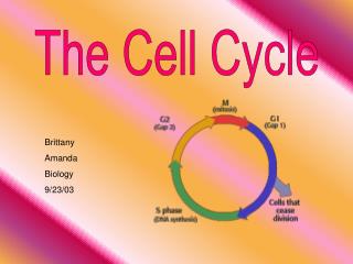
Overview of the Cell Cycle Source: www.els.net
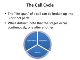
Let’s Start with G1!! • Fact: Most of the cells in our body are NOT replicating at any given point of time. • Instead, most cells in our body remain in the G0/G1 phase for their entire lifetime. • Most of the cells that go through the cell cycle on a regular basis are the adult stem cells in the bone marrow, skin, and the gut - continual cell division occurs to replenish the constantly dying cells in those organs.
G1 restriction point pRb: Tumor Suppressor Protein retinoblastoma E2F-DP: Transcribes genes whose products are used in DNA synthesis as well as for the genes for Cyclin E, Cyclin A, and Cdk2. Various growth factors and hormones activate Cdk4/6 & Cdk2 which phosphorylate pRb… Source: www.eurogene.org
DNA Damage Checkpoint [p21 is a CDKI which binds Cdk2 and Cdk4/6] Source: www.eurogene.org
Triggering the S phase • The newly formed CycA-Cdk2 complexes help “trigger” pre-replication complexes assembled on the DNA by causing the assembly of DNA polymerase, thus initiating the S-phase. Source: www.med.upenn.org
More CycA-Cdk2 S-phase Regulation • CycA-Cdk2 phosphorylates E2F to inactivate its function as a transcription factor. • CycA-Cdk2 also keeps the pRb protein inactive during the S phase. • CycA-Cdk2 also phosphorylates Cdc6 (part of the pre-replicative complex) and thus marks it for degradation – thus ensuring that re-replication does not occur.
G2/M transition (Cdc2=Cdk1) • During the G2 phase of the cell cycle, inactive cyclin B1/Cdc2 complexes accumulate in mammalian cells due to inhibitory phosphorylation of Cdc2 on Thr-14 and Tyr-15 by the Wee1 and Myt1 kinases. • CyclinB1/Cdc2 activation requires dephosphorylation on Thr-14 and Tyr-15 by the phosphatase Cdc25C, and phosphorylation on Thr-161 by CAK3,4.
Activation of Cdc25C • The mammalian Plx1 homolog Plk1 phosphorylates Cdc25C. • Active cyclin B1/Cdc2 activates Cdc25C by phosphorylating the N-terminal region of Cdc25C in an autocatalytic loop. Thus, it takes only a small amount of active cyclin B1/Cdc2 for its concentration to rapidly explode.
G2 Checkpoint Activation • In response to genotoxic stress such as Ionizing Radiation or Ultra Violet light, the ATM/ATR signaling pathway is activated. These two kinases are part of the phosphoinositide-3 kinase (PI-3K) family. • ATM phosphorylates and activates Chk2; ATR, Chk1. • Activated Chk1 and Chk2 phosphorylate Cdc25C on Ser-216, generating a consensus binding site for 14-3-3 proteins. • Cdc25C is then exported out of the nucleus and is sequestered in the cytoplasm by the 14-3-3 proteins. • This leads to G2 cell cycle arrest due to the inhibition of the ability of Cdc25C to activate cyclin B1/Cdc2. • Cdc25C phosphorylation happens at a different site than when it is done by Plk1, as mentioned in the previous slide. Source: www.eurekah.com
Mitosis Stage 1: Prophase • The replicated chromosomes condense to become very thick and dense. • Microtubules start sprouting from the centrosomes which had also replicated during the S-phase of the cell cycle. • The centrosomes start to move apart.
Mitosis Stage 2: Prometaphase • The nuclear envelope abruptly breaks down. • Microtubules start attaching to the kinetochores of the chromosomes. • Centrosomes go towards opposite poles; chromosomes start moving towards center. Source: www.sparknotes.com
Mitosis Stage 3: Metaphase • The chromosomes align at the metaphase plate. • The metaphase checkpoint monitors the attachment of the mitotic spindle to kinetochores and the tension generated by mitotic spindle attachment. In the presence of even a single unattached kinetochore, the metaphase checkpoint halts the separation of sister chromatids and thereby provides additional time for spindle attachment. Source: http://www.stanford.edu/group/fanglab/science/research_cycle.html
Mitosis Stage 4: Anaphase • The breakdown of the cohesin protein complex that holds the sister chromatids together is done by the Anaphase Promoting Complex (APC). • The APC works by degrading a protein that is binding a proteolytic enzyme that cleaves the cohesin when it is no longer held inactive by the now-degraded protein.
Mitosis Stage 6: Cytokinesis • Cleavage occurs by the contraction of a thin ring of actin filaments that form the contractile ring. • If the ring is not positioned at the center of the cell, an asymmetrical division takes place.
Importance of Cell Size • As the cell grows in size, more and more ribosomes are produced. So, more and more cyclins are produced, and so more and more Cyclin-dependent Kinase complexes form (e.g. CyclinA-Cdk2 going into S; CyclinB-Cdk1 going into M)…since the nucleus size doesn’t increase, these become more and more concentrated -> more active->carry out the transition into the next cell cycle phase.