Download

1 / 52
520 likes | 547 Views
Explore the importance of Vitamin A in normal visual function, cell growth, immunity, and more. Learn about sources, absorption, recommended intake, and storage. Dive into its role in vision, growth, and reproduction.

E N D
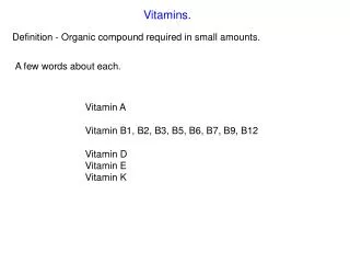
Vitamin A Chemistry, Functions and Deficiency 1
Introduction • Vitamin A is a fat soluble Vitamin and is Important for • Normal Visual function • Maintenance of cell function and growth • Epithelial integrity • Red blood cell production • Immunity and • Reproduction. • Vitamin A deficiency (VAD) is a major nutritional concern in poor societies, especially in lower income countries like Pakistan
Lipid soluble vitamins - commonfeatures • They cannot be synthesized by thebody • Supplied by thediet and Absorbedalong withfats. • Transportedby LP& Specific bindingproteins • If Extra, are stored in liver andadiposetissue. • Excess consumption leads to accumulation andcan be toxic.
Two groups of compounds have Vitamin A activity. • Retinoids • Carotenoids 2
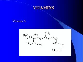
Vitamin A, in the strictest sense, refers to retinol. However, theoxidized metabolites,retinaldehyde andretinoic acid,are also biologically active compounds. The term Retinoids includes all molecules (including synthetic retinol. molecules) that are chemically related to 3
More than 600 carotenoids innature,and approximately 50 of these can bemetabolized tovit. A. • The α-, β-, and γ-carotenes are quantitatively the most important provitamin A carotenoids. • β-Caroteneis the most prevalent carotenoid in the food supply that has provitamin A activity. • In humans,carotenoids are absorbed intact and are stored in liver and fat. 4
Liver, fish,and eggs areexcellent food sources forpreformed vitamin A. Vegetable sources of provitamin A carotenoids include dark green and deeply colored fruits and vegetables. • • Moderate enhances uptakein cooking of vegetables carotenoid release for the gut. • 7
Recommended Dietary Allowance (RDA) Vitamin A for Adults • Women: 700 µg or 2,330 IU µg • Men: 900 µg or 3,000 IU • UL Men or Women: 3,000 µg or 10,000 IU
Attaching group β – Ionone ring Unsaturated Isoprenoid side chain (all trans) 5
•Site of β cleavage of carotene is shown by * •One molecule of β caroteneyields two molecules of vit A retinol •6 μg of β carotene is equivalent to 1μ g of preformed retinol. •The total amount of vitamin A in foods is therefore expressed asmicrograms of retinol equivalents (RE). 6
Vitamin A, in animal sources is in theform of Retinyl esters. • It is hydrolyzed to retinol and fatty acid by pancreatic hydrolases. • Absorption requires the presence of bile salts. • In intestinal cells, retinol is esterified back and secreted with chylomicrons. • Carotenoid absorption is also aided by some fat in meal. • 90% of retinoidscan beabsorbed 8
β-Carotene is cleaved in theintestinal mucosa by carotene dioxygenase. • It yieldsretinal, whichis reducedtoretinol. • Retinol is esterified andsecretedin chylomicrontogether with esters formedfrom dietary retinol. 9
Absorption of Carotenoids Absorbed intact, absorption rate much lower Intestinal cells can convert carotenoids toretinoids. • It is passed along with fat through the lymphatic system into bloodstream. • Absorption increases if taken withfat. • Approximately 80% isabsorbed • Vitamin A which is not absorbed is excreted within 1 or 2 days in feces.
Transport • Chylomicrons from intestinal cells tothe liver • From liver to target tissue as retinolvia retinol-binding protein (RBP) • The retinol-binding protein complex interacts with a second protein, Transthyretin.
This trimolecular complex functions to prevent vitamin A • Frombeing filtered by the kidney glomerulus • To protect the body against the toxicity of retinol and • To allow retinol to be taken up by specific cell-surface receptors that recognize retinol-binding protein. 10
Storage • The liver has enormous capacity to store in the formofretinolpalmitate. • Under normal conditions a well-fed person has sufficient Vitamin A reserves to meet his need for 6 to 9months or more.
Excretion of VitaminA • Not readilyexcreted • Aging increase risk of toxicity because excretion isimpaired
Essential for vision Immuneresponse, Bone growthReproduction • • • • • Maintenance of the surface linings of the eyes, epithelial cell growth and repair, and theepithelial integrity of therespiratory, urinary, and intestinal tracts. Vitamin A is also important for embryonic development and the regulation of adult genes. •
Functions of VitaminA • Vision: Vitamin A is a component of the visualpigment rhodopsin. Retinal is bound to the proteinopsin. • Growth: Vitamin A deficiency causes loss of appetite. Slow bone growth. AffectsCNS. • Reproduction: Retinol and retinal are essential fornormal reproduction • Maintenance of epithelial cells: Essential for normal differentiation of epithelial tissues and mucussecretion
Role inVision • VisualCycle(Wald’sVisualCycle) • A process by which light fallingon the retina of theeye is converted to an electricalsignal • The optic nerve carries the electrical signal to thebrain (nerveimpulse) • The brain processes the signal into animage
Role inVision • Retina is a light-sensitive layer of cells ofthe eye • Retina consists of: Rod and cone cells (photosensitive cells) • Rod cellsprocess black and white image • Cone cells process colorimage
Role of Vitamin A inVision • Normal vision depends on the retina and onvitaminA • In the retina, vitamin A in the form of Retinal binds to a protein called opsinto make Rhodopsin[11-cis –retinal- opsin] in rodcells • Rhodopsin is a light-sensitivepigments
Role of Vitamin A inVision • When stimulated by light, vitamin A isomerizes from its bent ‘cis’ form to a straighter ‘trans’ form and detaches fromopsin • The opsin molecule changes shape, which sends a signal to the brain via optic nerve and an image isformed • Most retinal released in this process is quickly converted to trans-retinol and then to cis-retinal, tobegin another cycle
Role of Vitamin A inVision • Dark Adaptationtime • Bright light depletes rhodopsin(photo bleaching) • Suddenshiftfrom bright to dim light, causes difficulty in seeing. • Rhodopsin is synthesized in few minutes and vision is improved in thedark
Role of Vitamin A inVision • The time required to synthesize rhodopsin in thedark is called dark adaptationtime • It is increased in vitamin Adeficiency
In the retina, retinaldehyde functions prosthetic group of the light-sensitive proteins, forming Rhodopsin(inrods) iodopsin (in cones). as the opsin and • Any one cone cell contains only one type of opsin, and is sensitive to only one color. The absorption of light by Rhodopsin causes isomerization of the retinaldehyde from 11-cis • • to all-trans, and a conformational change in opsin. This results in the release of retinaldehyde from the protein, and the initiationof anerve impulse. •
The formation of the initial excited form of Rhodopsin, bathorhodopsin, occurs within picoseconds of illumination. There are then a series of conformational changes leading to the formation metarhodopsin II, which initiates nucleotideamplification cascade nerveimpulse. of a guanine and then a
The final step is hydrolysis to release all- trans-retinaldehyde and opsin. Thekey toinitiation of the visualcycleis the availability of 11-cis-retinaldehyde, and hence vitamin A. In deficiency, both the time taken to adapt to darkness and the light are impaired. ability to see in poor
Vitamin A deficiency can result from inadequate intake, fat malabsorption, or liver disorders. Deficiency impairs immunity and hematopoiesis and causes skin rashes and typical ocular effects (e.g., xerophthalmia, night blindness). Diagnosis is based on typical ocular findings and low vitamin A levels. Treatment consists of vitamin A given orally or, if symptoms aresevere or malabsorption is the cause, parenterally.
Primary vitamin A deficiency • Prolonged dietary deprivation • Vegetarians, • Chronic alcoholics, • Toddlersand • Preschoolchildren.
Secondary Sprue, vitamin A deficiency o o o o o o o Cystic fibrosis, Pancreatic insufficiency, Duodenal bypass, Chronic diarrhea, Bile duct obstruction, Cirrhosis.
Bitot spots - Areas of abnormal squamous cell proliferation and keratinization of the conjunctiva can be seen in young children with VAD. Blindness due to retinal injury - Vitamin A has a major role in phototransduction. VAD leads to lackof visual pigments; this reduces the absorption of various wavelengths of light, resulting in blindness. a
Poor adaptation to darkness (nyctalopia), • which can lead tonight blindness, is an early symptom. • Xerophthalmiaresults from keratinization of theconjunctiva. • Keratomalacia- In advanced deficiency; the cornea becomes hazy and can develop erosions, which can lead to its destruction (
Increased susceptibility to infections- Keratinization of themucous membranes of respiratory tracts and urinary tract takes place, increasing the susceptibility to infections. Duringinfection the synthesis of retinol binding protein is reduced in response toinfection since it is a negative ‘Acute phase protein’, that results in decreased circulatory concentration of the vitamin deterioration of theimmune with further system.
Fatigue Anemia Diarrhea Respiratory infections Decreased Decreased Infertility growth rate bone development
Serum retinol level-Normal range is 28 to 86 μg/dL (1 to 3 µmol/L). The level decreases vitamin A deficiency. Serum RBP level Serum zinc level is useful because zinc deficiency interferes with RBP production. An iron panel is useful because iron in deficiency can affect the metabolism vitamin A. of
Albumin levels are indirect measures of vitamin A levels. Complete blood count (CBC) with differential if anemia, infection, or sepsis is a possibility. An electrolyte evaluation and liver function studies should be performed to nutritional and volume status. evaluate for 31
Effects 1. Night blindness (nyctalopia) Inability to see well in dim light easily when entering a dark space form bright light 2. Night blindness occurs when there is insufficient Vit A in the blood to quickly regenerate visual purple . 3. Alcoholic liver disease (cirrhosis) causes night blindness which is due to hepatic damage affecting Vit A release.
Effects • In the eye, the 1st symptom of Vit A deficiency include photophobia (sensitivity to bright light) • Inflammation of eyes and eyelids due to impaired functioning of lacrimal glands • Xerophthalmia (dry, inflamed and edematous cornea) • Keratomalcia – permanent blindness results when infection leads to ulceration and softening of cornea
Skin and mucous membrane changes • Keritinization of the epithelial tissues • Increase susceptibility to infections of all membranes, protected by mucous • Follicular hyperperatosis – The sebceous glands becomes clogged and skin takes on a gooseflesh like appearance.
Toxic effects • Drying and desquamation of skin • Anorexia • Loss of hair • Bone pain and fragility • Enlargement of liver and spleen