Download
1 / 33
330 likes | 354 Views
Dive into the intricate details of the heart's location, structure, and function. Learn about its walls, valves, and circulation to grasp its vital role in the body's overall functioning.

E N D
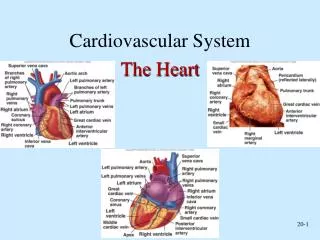
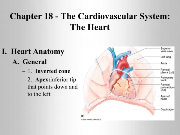
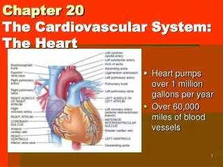
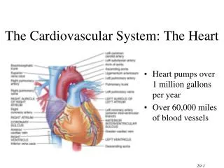
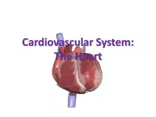
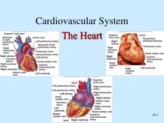
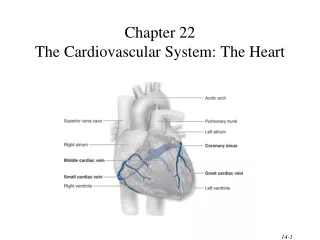
Heart Location • Heart is located in the mediastinum • area from the sternum to the vertebral column and between the lungs
Heart Orientation • Heart has 2 surfaces: anterior and inferior, and 2 borders: right and left • Apex - directed anteriorly, inferiorly and to the left • Base - directed posteriorly, superiorly and to the right
Pericardium • A dual layer membrane containing fluid • Prevents the heart from damage when beating (the surface of the sternum is rough) • Made up of two layers - visceral layer; up against heart muscle - parietal layer; outermost layer, touches lungs, sternum, and other organs
Layers of Heart Wall • Epicardium • visceral layer of serous pericardium • Myocardium • cardiac muscle layer is the bulk of the heart • Endocardium • chamber lining & valves
Right Atrium • Receives blood from 3 sources • superior vena cava, inferior vena cava and coronary sinus • Interatrial septum partitions the atria • Fossa ovalis is a remnant of the fetal foramen ovale • Tricuspid valve • Blood flows through into right ventricle • has three cusps composed of dense CT covered by endocardium
Right Ventricle • Forms most of anterior surface of heart • Papillary muscles are cone shaped trabeculae carneae (raised bundles of cardiac muscle) • Chordae tendineae: cords between valve cusps and papillary muscles • Interventricular septum: partitions ventricles • Pulmonary semilunar valve: blood flows into pulmonary trunk
Left Atrium • Forms most of the base of the heart • Receives blood from lungs - 4 pulmonary veins (2 right + 2 left) • Bicuspid valve: blood passes through into left ventricle • has two cusps • to remember names of this valve, try the pneumonic LAMB • Left Atrioventricular, Mitral, or Bicuspid valve
Left Ventricle • Forms the apex of heart • Chordae tendineae anchor bicuspid valve to papillary muscles (also has trabeculae carneae like right ventricle) • Aortic semilunar valve: • blood passes through valve into the ascending aorta • just above valve are the openings to the coronary arteries
Myocardial Thickness and Function • Ventricle walls are much thicker and stronger • right ventricle supplies blood to the lungs (little flow resistance) • left ventricle wall is the thickest to supply systemic circulation • Thickness of myocardium varies according to the function of the chamber • Atria are thin walled, deliver blood to adjacent ventricles
Thickness of Cardiac Walls Myocardium of left ventricle is much thicker than the right.
Atrioventricular Valves Close • A-V valves close preventing backflow of blood into atria • occurs when ventricles contract, pushing valve cusps closed, chordae tendinae are pulled taut and papillary muscles contract to pull cords and prevent cusps from everting
Atrioventricular Valves Open • A-V valves open and allow blood to flow from atria into ventricles when ventricular pressure is lower than atrial pressure • occurs when ventricles are relaxed, chordae tendineae are slack and papillary muscles are relaxed
Semilunar Valves • SL valves open with ventricular contraction • allow blood to flow into pulmonary trunk and aorta • SL valves close with ventricular relaxation • prevents blood from returning to ventricles, blood fills valve cusps, tightly closing the SL valves
Animation • The cardiac cycle
Blood Circulation • Two closed circuits, the systemic and pulmonary • Systemic circulation - left side of heart pumps blood through body - left ventricle pumps oxygenated blood into aorta - aorta branches into many arteries that travel to organs - arteries branch into many arterioles in tissue - arterioles branch into thin-walled capillaries for exchange of gases and nutrients - deoxygenated blood begins its return in venules - venules merge into veins and return to right atrium
Blood Circulation (cont.) Pulmonary circulation • right side of heart pumps deoxygenated blood to lungs • right ventricle pumps blood to pulmonary trunk • pulmonary trunk branches into pulmonary arteries • pulmonary arteries carry blood to lungs for exchange of gases • oxygenated blood returns to heart in pulmonary veins
Coronary Circulation • Coronary circulation is blood supply to the heart • Heart as a very active muscle needs lots of O2 • When the heart relaxes high pressure of blood in aorta pushes blood into coronary vessels • Many anastomoses • connections between arteries supplying blood to the same region, provide alternate routes if one artery becomes occluded
Autorhythmic Cells Cells fire spontaneously, act as pacemaker and form conduction system for the heart SA node cluster of cells in wall of Rt. Atria begins heart activity that spreads to both atria excitation spreads to AV node AV node in atrial septum, transmits signal to bundle of His AV bundle of His the connection between atria and ventricles divides into bundle branches & purkinje fibers, large diameter fibers that conduct signals quickly Conduction System of Heart
Animation • Conduction system of heart
Heart Sounds • Stethoscope • Sounds of heartbeat are from turbulence in blood flow caused by valve closure • first heart sound (lubb) is created with the closing of the atrioventricular valves • second heart sound (dupp) is created with the closing of semilunar valves
Risk Factors for Heart Disease • Risk factors in heart disease: • high stress • high blood pressure • cigarette smoking • obesity & lack of regular exercise. • Other factors include: • diabetes mellitus • genetic predisposition • male gender • high blood levels of fibrinogen • left ventricular hypertrophy
Exercise and the Heart • Sustained exercise increases oxygen demand in muscles. • Benefits of aerobic exercise (any activity that works large body muscles for at least 20 minutes, preferably 3-5 times per week) are; • increased cardiac output • increased HDL and decreased triglycerides • improved lung function • decreased blood pressure • weight control.