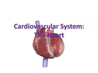
The heart Cardiovascular system
450 likes | 722 Views
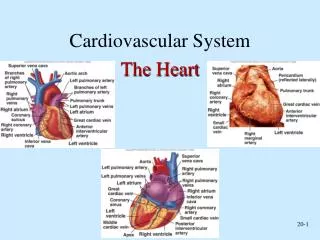
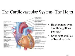
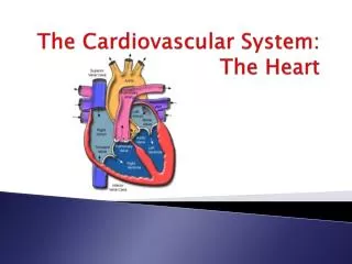
The heart Cardiovascular system . 1 Start with blood in the right atrium 2 Start with blood in the pulmonary vein 3. You pick!. Why is this picture deceiving?. Capillary beds of lungs where gas exchange occurs. Pulmonary Circuit. Pulmonary veins. Pulmonary arteries. Aorta and branches.

The heart Cardiovascular system
E N D
Presentation Transcript
1 Start with blood in the right atrium 2 Start with blood in the pulmonary vein 3. You pick!
Why is this picture deceiving? Capillary beds of lungs where gas exchange occurs Pulmonary Circuit Pulmonary veins Pulmonary arteries Aorta and branches Venae cavae Left atrium Left ventricle Right atrium Heart Right ventricle Systemic Circuit Oxygen-rich, CO2-poor blood Capillary beds of all body tissues where gas exchange occurs Oxygen-poor, CO2-rich blood Figure 18.5
Heart AnatomyYou know this… • Approximately the size of a fist • Location, location, location • In the mediastinum between second rib and fifth intercostal space • On the superior surface of diaphragm • Two-thirds to the left of the midsternal line • Enclosed in pericardium, a double-walled sac
Midsternal line 2nd rib Sternum Diaphragm Point of maximal intensity (PMI) (a) Figure 18.1a
Aorta Superior vena cava Parietal pleura (cut) Pulmonary trunk Left lung Pericardium (cut) Apex of heart Diaphragm (c) Figure 18.1c
Pericardium • Superficial fibrous pericardium • Protects, anchors, and prevents overfilling
Pericardium • Deep two-layered serous pericardium • See notes, October, 2011
Pulmonary trunk Fibrous pericardium Parietal layer of serous pericardium Pericardium Pericardial cavity Myocardium Epicardium (visceral layer of serous pericardium) Heart wall Myocardium Endocardium Heart chamber Figure 18.2
Layers of the Heart Wall • Epicardium—visceral pericardium -(layer of the serous pericardium) • Myocardium • Spiral bundles of cardiac muscle cells • Endocardium is continuous with endothelial lining of blood vessels
Pulmonary trunk Fibrous pericardium Parietal layer of serous pericardium Pericardium Pericardial cavity Myocardium Epicardium (visceral layer of serous pericardium) Heart wall Myocardium Endocardium Heart chamber Figure 18.2
Cardiac muscle bundles Figure 18.3
Chambers • Four chambers • Two atria • Separated internally by the septum
Chambers • Two ventricles • Separated by the interventricular septum • Anterior and posterior interventricular sulci mark the position of the septum externally
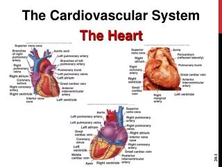
Left common carotid artery Brachiocephalic trunk Left subclavian artery Superior vena cava Aortic arch Ligamentum arteriosum Right pulmonary artery Left pulmonary artery Ascending aorta Left pulmonary veins Pulmonary trunk Right pulmonary veins Auricle of left atrium Circumflex artery Right atrium Left coronary artery (in coronary sulcus) Right coronary artery (in coronary sulcus) Anterior cardiac vein Left ventricle Right ventricle Great cardiac vein Right marginal artery Anterior interventricular artery (in anterior interventricular sulcus) Small cardiac vein Inferior vena cava Apex (b) Anterior view Figure 18.4b
Atria: The Receiving Chambers • Vessels entering right atrium • Superior vena cava • Inferior vena cava • Coronary sinus • Vessels entering left atrium • Right and left pulmonary veins
Ventricles: The Discharging Chambers • Papillary muscles project into the ventricular cavities • Vessel leaving the right ventricle • Pulmonary trunk • Vessel leaving the left ventricle • Aorta
Aorta Left pulmonary artery Superior vena cava Left atrium Right pulmonary artery Left pulmonary veins Pulmonary trunk Right atrium Mitral (bicuspid) valve Right pulmonary veins Fossa ovalis Aortic valve Pectinate muscles Pulmonary valve Tricuspid valve Left ventricle Papillary muscle Right ventricle Interventricular septum Chordae tendineae Trabeculae carneae Epicardium Inferior vena cava Myocardium Endocardium (e) Frontal section Figure 18.4e
Pathway of Blood Through the Heart • The heart is two side-by-side pumps • Right side is the pump for the pulmonary circuit • Vessels that carry blood to and from the lungs • Left side is the pump for the systemic circuit • Vessels that carry the blood to and from all body tissues
Capillary beds of lungs where gas exchange occurs Pulmonary Circuit Pulmonary veins Pulmonary arteries Aorta and branches Venae cavae Left atrium Left ventricle Right atrium Heart Right ventricle Systemic Circuit Oxygen-rich, CO2-poor blood Capillary beds of all body tissues where gas exchange occurs Oxygen-poor, CO2-rich blood Figure 18.5
Pathway of Blood Through the Heart • Right atrium tricuspid valve right ventricle • Right ventricle pulmonary semilunar valve pulmonary trunk pulmonary arteries lungs PLAY Animation: Rotatable heart (sectioned)
Pathway of Blood Through the Heart • Lungs pulmonary veins left atrium • Left atrium bicuspid valve left ventricle • Left ventricle aortic semilunar valve aorta • Aorta systemic circulation PLAY Animation: Rotatable heart (sectioned)
Pathway of Blood Through the HeartFun facts • Equal V of blood are pumped through both circuits • Pulmonary circuit is a short, low-pressure
Pathway of Blood Through the Heart • Systemic circuit blood encounters much more resistance in the long pathways • Anatomy of the ventricles reflects these differences
Left ventricle Right ventricle Interventricular septum Figure 18.6
Coronary Circulation • The functional blood supply to the heart muscle itself • Arterial supply varies person to person! • Collateral routes provide additional routes for blood delivery
Coronary Circulation • Arteries • Right – marginal • left coronary (in atrioventricular groove), circumflex, and anterior interventricular arteries • Veins • Small cardiac, anterior cardiac, and great cardiac veins
Aorta Pulmonary trunk Superior vena cava Left atrium Anastomosis (junction of vessels) Left coronary artery Right atrium Circumflex artery Right coronary artery Left ventricle Right ventricle Anterior interventricular artery Right marginal artery Posterior interventricular artery (a) The major coronary arteries Figure 18.7a
Superior vena cava Great cardiac vein Anterior cardiac veins Coronary sinus Small cardiac vein Middle cardiac vein (b) The major cardiac veins Figure 18.7b
Aorta Superior vena cava Left pulmonary artery Right pulmonary artery Right pulmonary veins Left pulmonary veins Auricle of left atrium Right atrium Left atrium Inferior vena cava Great cardiac vein Coronary sinus Right coronary artery (in coronary sulcus) Posterior vein of left ventricle Posterior interventricular artery (in posterior interventricular sulcus) Left ventricle Apex Middle cardiac vein Right ventricle (d) Posterior surface view Figure 18.4d
Homeostatic Imbalances • Angina pectoris • Thoracic pain caused by a fleeting deficiency in blood delivery to the myocardium • Cells are weakened
Homeostatic Imbalances • Myocardial infarction (heart attack) • Prolonged coronary blockage • Areas of cell death are repaired with noncontractile scar tissue
Heart Valves • Ensure unidirectional blood flow through the heart • Atrioventricular (AV) valves • Prevent backflow into the atria when ventricles contract • Tricuspid valve (right) • Mitral valve (left) • Chordae tendineae anchor AV valve cusps to papillary muscles
Heart Valves • Semilunar (SL) valves • Prevent backflow into the ventricles when ventricles relax • Aortic semilunar valve • Pulmonary semilunar valve
Pulmonary valve Myocardium Aortic valve Tricuspid (right atrioventricular) valve Area of cutaway Mitral valve Tricuspid valve Mitral (left atrioventricular) valve Myocardium Tricuspid (right atrioventricular) valve Aortic valve Mitral (left atrioventricular) valve Pulmonary valve Aortic valve Pulmonary valve Aortic valve Pulmonary valve Area of cutaway (b) Fibrous skeleton Mitral valve Tricuspid valve (a) Anterior Figure 18.8a
Myocardium Tricuspid (right atrioventricular) valve Mitral (left atrioventricular) valve Aortic valve Pulmonary valve Pulmonary valve Aortic valve Area of cutaway (b) Mitral valve Tricuspid valve Figure 18.8b
Pulmonary valve Aortic valve Area of cutaway Mitral valve Tricuspid valve Chordae tendineae attached to tricuspid valve flap Papillary muscle (c) Figure 18.8c
Opening of inferior vena cava Mitral valve Chordae tendineae Tricuspid valve Myocardium of right ventricle Myocardium of left ventricle Pulmonary valve Aortic valve Area of cutaway Papillary muscles Mitral valve Interventricular septum Tricuspid valve (d) Figure 18.8d
1 Blood returning to the heart fills atria, putting pressure against atrioventricular valves; atrioventricular valves are forced open. Direction of blood flow Atrium Cusp of atrioventricular valve (open) 2 As ventricles fill, atrioventricular valve flaps hang limply into ventricles. Chordae tendineae 3 Atria contract, forcing additional blood into ventricles. Papillary muscle Ventricle (a) AV valves open; atrial pressure greater than ventricular pressure Atrium 1 Ventricles contract, forcing blood against atrioventricular valve cusps. Cusps of atrioventricular valve (closed) 2 Atrioventricular valves close. Blood in ventricle 3 Papillary muscles contract and chordae tendineae tighten, preventing valve flaps from everting into atria. (b) AV valves closed; atrial pressure less than ventricular pressure Figure 18.9
Aorta Pulmonary trunk As ventricles contract and intraventricular pressure rises, blood is pushed up against semilunar valves, forcing them open. (a) Semilunar valves open As ventricles relax and intraventricular pressure falls, blood flows back from arteries, filling the cusps of semilunar valves and forcing them to close. (b) Semilunar valves closed Figure 18.10
Microscopic Anatomy of Cardiac Muscle • Cardiac muscle cells are striated, short, fat, branched, and interconnected • Connective tissue matrix (endomysium) connects to the fibrous skeleton • T tubules are wide but less numerous; SR is simpler than in skeletal muscle • Numerous large mitochondria (25–35% of cell volume)
Nucleus Intercalated discs Cardiac muscle cell Gap junctions Desmosomes (a) Figure 18.11a
Microscopic Anatomy of Cardiac Muscle • Intercalated discs: junctions between cells anchor cardiac cells • Desmosomes prevent cells from separating during contraction • Gap junctions allow ions to pass; electrically couple adjacent cells • Heart muscle behaves as a functional syncytium
Cardiac muscle cell Mitochondrion Intercalated disc Nucleus T tubule Mitochondrion Sarcoplasmic reticulum Z disc Nucleus Sarcolemma I band I band A band (b) Figure 18.11b