Download
1 / 30
300 likes | 423 Views
Chapt 19: Ribosomes and Transfer RNA. Student learning outcomes Describe basic structure of the ribosome, relationship of two subunits - catalytic roles of RNA Describe basic structure of tRNA Explain how amino acyl tRNA synthetases provide second code – insert correct amino acid on tRNA

E N D
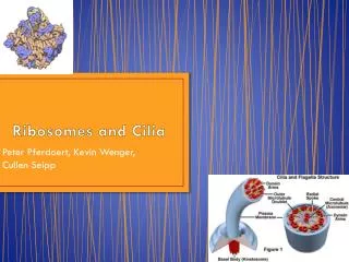
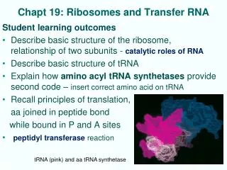
Chapt 19: Ribosomes and Transfer RNA Student learning outcomes • Describe basic structure of the ribosome, relationship of two subunits - catalytic roles of RNA • Describe basic structure of tRNA • Explain how amino acyl tRNA synthetases provide second code – insert correct amino acid on tRNA • Recall principles of translation, aa joined in peptide bond while bound in P and A sites • peptidyl transferase reaction tRNA (pink) and aa tRNA synthetase
Appreciate Nobel Prizes for 2010 for ribosome structure and function: • Tom Steitz 50S ribosome structure Haloarcula • Venkatraman Ramakrishnan – 30S structure Thermus thermophilus • Ada Yonath – 30S structure Thermus thermophilus, started work crystallography Geobacillus, Haloarcula • Important Figures: 1, 2, 3*, 7, 8, 14, 15, 18, 20, 22, 24, 25, 26*, 28*, 31 • Review problems: 1, 3, 4, 5, 6, 8, 13, 14, 15, 19; AQ 1, 2
Bacterial Ribosome Composition • E. coli ribosome 70S • 30 subunit: • 16S rRNA • 21 proteins (S1 – S21) • 50S subunit: • 5S rRNA • 23S rRNA • 34 proteins (L1 – L34) Eukaryotic organelle ribosomes are similar, smaller Fig. 3.16
19.1 Bacterial Ribosomes 30S - small subunit decodes mRNA 50S –large subunit links amino acids together through peptide bonds • Eukaryotic cytoplasmic ribosomes: • Larger (80S,- 40S, 60S • more RNAs, more proteins • 28S, 18S, 5.8S, 5S Fig. 4 Ribosome with 3 tRNAs in A (aminoacyl), P (peptidyl) and E (exit) sites
Brief recall Protein synthesis • Prokaryotes: polycistronic • mRNA binds 30S subunit at ribosome binding site • 1st tRNA is fmet (N-formyl-methionine) in P site • Lots of protein factors (IF, EF), GTP help • 50S subunit binds • 2nd tRNA binds to A site; peptide bond forms • Translocation of tRNA-peptide to E site Eukaryotes:monocistronic • Ribosomes bind CAP, scan to find 1st AUG • 1st tRNA is met, not fmet
Elongation: peptidyl transferase of 50Sjoins amino acids in peptide bond GTP and many protein factors are required; Incoming aa-tRNA receives growing polypeptide chain Translocation and exit of empty tRNA Fig. 3.19
Fine Structure of 70S Ribosome • BacterialThermus thermophilus crystal structure: 70S ribosome with mRNA analog, 3 tRNAs : • Positions, tertiary structures of all 3 rRNAs, most proteins • Shapes and locations of tRNAs in A, P, and E sites • Binding sites for tRNAs in ribosome are rRNA, not protein • Contacts between subunits are mostly rRNA • Anticodons of tRNAs in A and P sites approach each other closely enough to base-pair with adjacent codons bound to 30S subunit as mRNA kinks 45° (Fig. 2)
Fig. 19.1 Thermus thermophilus a-d rotated versions; 30S front in a e, top with 50S top; f, g individual 50S, 30S 16S rRNA cyan 23S rRNA gray 5S RNA dark blue tRNA gold, orange
Fig. 2 tRNA bound to codons on ribosome Fig. 3 structure of ribosome showing tRNAs bound at interface of subunits
Ribosomal proteins identified by 2D gel electrophoresis • More sensitive than 1D: • 1st dimension pH 8.6, 8% acrylamide gel • 2nd dimension, pH 4.6 18% acrylamide • Also cloned genes and purifed proteins Fig. 5 E. coli proteins
Ribosome Assembly • E. coli assembly with purified proteins in vitro : • 30S ribosomal subunit begins with 16S rRNA • Proteins join sequentially and cooperatively • Proteins added early in process help later proteins to bind to growing particle Fig. 20, thick arrows strong facilitating, thin weaker
Fine Structure of 30S Subunit • Consensus sequences of 16S rRNA led to secondary structure • X-ray crystallography studies confirmed • 30S subunit - extensively base-paired 16S rRNA shape essentially outlines particle • X-ray crystallography confirmed locations of 30S ribosomal proteins • Three major domains Fig. 8 T. thermophilus 16S
Crystal structure of T. thermophilus ribosome 30S shows rRNA domains stereo Fig. 9 rRNA domains: H = head; N = neck; B = beak; Sh = shoulder; P = platform; Bo = body; Sp = spur
30S Subunit binds antibiotics, initiation factors • 2 roles of 30S ribosomal subunit: • Facilitates proper decoding between codons and aminoacyl-tRNA anticodons • Also participates in translocation • Crystal structures of 30S subunits with interfering antibiotics sheds light on translocation and decoding • Spectinomycin – interferes with translocation • Streptomycin – error rate increases • Paromomycin – decreases accuracy of translation (A site) Antibiotic-resistant mutants can arise from altered ribosomal proteins (S12) 30S binds initiation factors (IF)
Fine Structure of 50S Subunit - Steitz • Crystal structure to 2.4 Å • Relatively few proteins at interface between ribosomal subunits • No proteins within 18 Å of peptidyl transferase active center (tagged with transition state analog) • 2’-OH group of tRNA in P site forms H bond to amino group of aminoacyl-tRNA in A site Fig.16 50S of Archaeon Haloarcula; green is peptidyl transferase region; yellow proteins
Role of 2‘-OH of tRNA • 2’-OH group of tRNA in P site: Forms H-bond to amino group of aminoacyl-tRNA in A site Helps catalyze peptidyl transferase reaction • Removal of 2’-OH group eliminates peptidyl transferase activity (Fig. 19) Fig. 18 Peptide bond involves Nucleophilic attack by aa in A site to COO- joined to tRNA in P site; Amino acid joined to 3’-OH of tRNA
50S Exit Tunnel Exit tunnel through 50S subunit • Just wide enough to allow protein a-helix to pass • Walls of tunnel made of RNA • Hydrophilicity likely to allow exposed hydrophobic side chains of nascent polypeptide to slide easily (not bind) Fig. 20
Polysomes • mRNAs translated by > one ribosome at at time • Polysome: structure in which many ribosomes translate mRNA in tandem • Eukaryotic polysomes are found in cytoplasm • In Prokaryotes, transcription of gene and translation of resulting mRNA occur simultaneously • [Many polysomes associated with active gene] Fig. 21 polysomes in bacteria; transcirption and translation simultaneously
19.2 Transfer RNA • Adaptor molecule (proposed by Crick, 1958) as mediator between string of nucleotides in DNA or RNA and string of amino acids in protein • 3 nucleotides could pair with nucleotides in codons Fig. 24 tRNA molecule
Discovery of tRNA 3’ CCA-aa 5’ • Small, independent of ribosome • Could be charged with amino acid: covalently joined in process requiring ATP • Charged species transfers amino acid to growing polypeptide: • Amino end of 2nd amino acid attacks COO- of first aa (which COO- is joined through tRNA)
tRNA Structure:cloverleaf • common secondary structure • 4 base-paired stems define 3 stem-loops • D loop - dihydrouracil • Anticodon loop • T loop (TYC sequence – Y = pseudouridine) • Acceptor stem - site amino acids are added
tRNA Shape • Common 3-D shape resembling inverted L • Maximizes stability by lining up base pairs: • D stem to anticodon stem • T stem to acceptor stem • Anticodon protrudes from side of loop • Anticodon shape base-pairs with mRNA codon
Modified Nucleosides occur in tRNA Fig. 25 Modifications occur during processing of tRNA; many enzymes required
Amino acyl tRNA synthases add amino acids – second genetic codeStructure of tRNA-amino acid • Amino acid covalently joined to specific tRNA at terminal 3’-CCA sequence • Amino terminal end of aa-2nd tRNA attacks COO- of 1st aa-tRNA to form peptide bond Fig. 17.1
Charging tRNA with amino acid: requires ATP, aminoacyl tRNA synthase Fig. 17.2
Changing amino acid chemically after charging results in insertion of wrong amino acid Ribosome recognizes tRNA not the amino acid Fig. 28 chemically altered cys in cys-tRNA resulted in incorrect amino acid ala being inserted in synthetic mRNA
tRNA Acceptor Stem and anticodon are important for aa tRNA synthase • Biochemical and genetic experiments demonstrated: • acceptorstem recognized in tRNA by cognate aa-tRNA synthetase • Changing one base pair in acceptor stem can change charging specificity • Second genetic code: charging correct amino acid • Anticodon, is also important element in charging specificity
Structures of Synthetase-tRNA Complexes Interactions differ between 2 classes of aminoacyl-tRNAsynthetases: opposite sides • Class I approach D loop, minor groove of acceptor stem • E.g. GlnRS-tRNAgln • B) class 2 bind variable region, major groove of acceptor stem • E.g. AspRS-tRNAasp Fig. 30
aa- tRNA synthetases also proofread and edit aa selectivity controlled by double-sieve mechanism • 1st sieve is coarse, excluding amino acids too big • Active site for activation of amino acids is just big enough for cognate amino acid, not larger amino acids • 2nd sieve is fine, degrades too small aminoacyl-AMPs • Editing site admits small aminoacyl-AMPs and hydrolyzes • Cognate aminoacyl-AMP is too big to fit editing site • Enzyme transfers activated amino acid to cognate tRNA
Review questions 3. What parts of tRNA interact with 30S? With 50S? 4,5. Why is it important that the anticodons in A & P sites, and that tRNA acceptor stems in A & P sites approach each other closely? 14. Draw cloverleaf tRNA structure and draw important structural elements. Draw the charged tRNA with an amino acid; diagram how one aa-tRNA is joined to the growing peptide chain (remember 5’, 3’).