Download
1 / 14

E N D
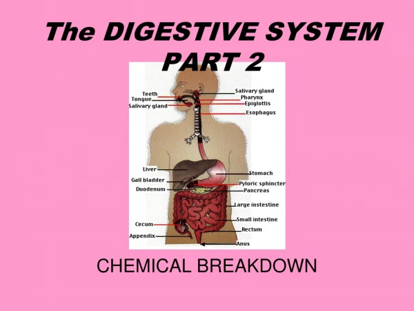
1. UNIT 8 The Digestive System Part 2 of 2 (Chapter 23) Functional Anatomy - The Liver and Gallbladder
Functional Anatomy - The Pancreas
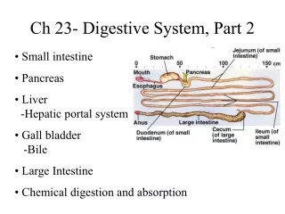
Functional Anatomy - The Large Intestine
Digestive Physiology: Motility, Secretion, Digestion, and Absorption
Digestive Pathologies and Disorders
(7th edition)
2. Functional Anatomy - The Liver and Gallbladder Functions of the Liver
largest gland in the body
performs over 500 functions, including many metabolic functions
converts glucose into glycogen
detoxifies many poisons and drugs (e.g. alcohol)
hepatocytes (liver cells) produce bile, an emulsifier of fats - it turns large droplets of fat into many smaller droplets, thus increasing surface area and promoting more efficient fat digestion (more surface area for enzymes to act upon); remember that small objects have more relative surface area (higher surface area to volume ratios)
Gross Anatomy of the Liver (fig. 23.23)
4 lobes: right lobe, left lobe, caudate lobe, quadrate lobe
ligamentum venosum (remnant of fetal circulation; in the fetus it served as a liver bypass), ligamentum teres (remnant of the fetal umbilical vein), and falciform �ligament� (a mesentery)
hepatic portal vein carries blood from digestive tract to the liver
bile is stored and concentrated in the gallbladder (note: the gallbladder does not make bile; that is the job of the liver)
(7th edition)
3. Functional Anatomy - The Liver and Gallbladder Microscopic Anatomy of the Liver (fig. 23.24)
liver lobules containing hepatocytes (=liver cells) radiate out from a central vein
portal triad = portal arteriole + portal venule + a bile duct
Kupffer cells - destroy bacteria and other foreign particles
Bile Flow (fig. 23.20)
when the hepatopancreatic sphincter is relaxed: bile flows from the right and left hepatic ducts of the liver into the common hepatic duct, then into the (common) bile duct, out through the hepatopancreatic ampulla, and finally into the duodenum where it is needed
when the hepatopancreatic sphincter is contracted: bile backs up into the (common) bile duct and cystic duct, and finally the gallbladder, where it is stored
*Thus, the hepatopancreatic sphincter controls whether bile is secreted or stored; when no digestion is occurring the sphincter remains closed
(7th edition)
4. Functional Anatomy - The Pancreas Functions
both an exocrine gland (enzymes) and endocrine gland (hormones)
makes, stores, and secretes enzymes for digestion
produces the hormones glucagon and insulin to regulate blood glucose
Anatomy of the Pancreas (fig. 23.20)
head and tail; the head touches the duodenum and the tail connects to the spleen
main pancreatic duct and accessory pancreatic duct
hepatopancreatic ampulla and sphincter control release of pancreatic secretions
Microscopic Anatomy - pancreatic islets (islets of Langerhans) contain clusters of hormone secreting cells (secrete insulin and glucagon)
(7th edition)
5. Functional Anatomy - The Large Intestine Functions
small amount of digestion by bacteria
absorption of water and electrolytes to form feces
Gross Anatomy of the Large Intestine (fig. 23.29)
cecum - first portion of the large intestine; connects to the ileum at the ileocecal valve
vermiform appendix - �worm-shaped�; has some lymphatic function
colon - ascending, transverse, descending, and sigmoid segments
rectum - located in the pelvic region
anal canal (fig. 23.29b)- internal anal sphincter (smooth muscle) and external anal sphincter (skeletal muscle); remember that you have voluntary control over skeletal muscle but not smooth muscle; thus, you can voluntarily contract the external anal sphincter, but not the internal anal sphincter
teniae coli - maintain constant muscle tone to constrict the large intestine and form pouches called haustrae
(7th edition)
6. Functional Anatomy - The Large Intestine Microscopic Anatomy of the Large Intestine
unlike the small intestine, there is an absence of villi and microvilli; because digestion and absorption are not the primary functions of the large intestine, it does not need the added surface area provided by villi and microvilli
like the small intestine, the lining is composed of simple columnar epithelium
(7th edition)
7. Digestive Physiology: MOTILITY two purposes:
moving food from mouth to anus
mechanically mixing food to break it down into smaller pieces, to increase surface area for exposure to digestive enzymes
nerves (e.g. vagus nerve), hormones, and paracrines can alter motility; parasympathetic input from the vagus nerve increases motility (remember that we call the parasympathetic nervous system the �rest and digest� part)
smooth muscle of the GI tract is connected by gap junctions to create contracting segments
like cardiac muscle, autorhythmic cells spontaneously depolarize to cause contraction; in this case, contraction of smooth muscle in the wall of the GI tract
(7th edition)
8. Digestive Physiology: MOTILITY peristaltic contractions (fig. 23.3) = peristalsis; progressive waves of contraction move from one section of the GI tract to another; the ANS, hormones, and paracrines influence peristalsis in all regions of the GI tract
circular muscle contract just behind a bolus (mass of food)
this pushes the bolus forward into a receiving segment, where the circular muscles are relaxed
the receiving segment then pushes the mass forward, continuing the forward movement
segmental contractions (fig. 23.3) - segments of the small intestine contract and relax; alternating segmental contractions churn intestinal contents back and forth, mixing them and keeping them in contact with the absorptive epithelium
(7th edition)
9. Digestive Physiology: SECRETION typically, about 9 liters of fluid pass through an adult�s GI tract in one day; most is reabsorbed, and thus not lost to the external environment
fluid input = about 2.0 L from food and drink; 7.0 L from digestive secretions (e.g. saliva, bile, mucus, and various digestive enzymes)
fluid removal from GI tract = about 7.5 L absorbed from small intestine and 1.4 L absorbed from large intestine; 0.1 L excreted in the feces
some enzymes are secreted in an inactive form; for example, specialized cells in the stomach produce pepsinogen (inactive); the inactive pepsinogen does not damage the cells that produce it; later, upon encountering a lower pH environment, the pepsinogen is converted into the active form, pepsin
mucus is a viscous secretion - it forms a protective coating over the mucosa of the GI tract, and helps to lubricate the contents of the gut
mucous cells - secrete mucus in the stomach
goblet cells - part of the simple columnar epithelium of the intestine; secrete mucus in the small and large intestines
salivary glands - secrete mucus in the saliva of the mouth
(7th edition)
10. Digestive Physiology: DIGESTION and ABSORPTION digestion of macromolecules (carbohydrates, proteins, and fats) is accomplished by a combination of mechanical and chemical processes
chewing and churning (mechanical) create smaller pieces of food, which expose more surface area to digestive enzymes; segmentation (mechanical) results in food mixing
bile from the liver creates droplets of lipids (fats) with greater surface area; bile is an emulsifier of fats
the optimum pH for various digestive enzymes reflects the location where they are most active; enzymes in the stomach have an acidic optimum pH, whereas enzymes in the small intestine work best at alkaline (basic) pH
most nutrient absorption takes place in the small intestine; some additional absorption of water and ions also occurs in the large intestine
(7th edition)
11. Digestive Physiology: DIGESTION and ABSORPTION carbohydrate digestion: (fig. 23.33)
most carbohydrates are ingested in the form of starch (a polysaccharide) and disaccharides, such as sucrose (�table sugar�), lactose (milk sugar), and maltose; other dietary carbohydrates include monosaccharides (�simple sugars�) such as glucose and fructose, and the polysaccharides glycogen and cellulose (�fiber�)
we are unable to digest cellulose, also known as fiber, because we lack the necessary enzymes
the enzyme amylase breaks down starch into smaller glucose chains and into the disaccharide maltose
other enzymes (e.g. maltase, lactase, and sucrase) break down their corresponding disaccharides into monosaccharides; maltase breaks maltose down into 2 glucose molecules, lactase breaks lactose down into glucose and galactose, and sucrase breaks sucrose down into glucose and fructose; the disaccharides must be broken down into monosaccharides before they can be absorbed across the epithelium of the GI tract
(7th edition)
12. Digestive Physiology: DIGESTION and ABSORPTION protein digestion: (fig. 23.33)
plant protein is the least digestible, whereas animal protein is the most digestible (egg protein is the best, with 85-90% in a form that can be digested and absorbed)
two broad groups of protein enzymes: endopeptidases and exopeptidases; endopeptidases attack peptide bonds in the interior of the amino acid chain, whereas exopeptidases chop off single amino acids from the ends of the amino acid chains
(7th edition)
13. Digestive Physiology: DIGESTION and ABSORPTION lipid digestion: (fig. 23.33)
on average, about 90% of our ingested fat comes from triglycerides, because they are the primary form found in both plants and animals
other lipid molecules in our diet include cholesterol, phospholipids, and fat-soluble vitamins
most lipids are not water-soluble; therefore, they appear as clumps in the chyme solution of the digestive tract; thus, they must be first broken down into smaller particles (via bile) before digestion can proceed
enzymatic fat digestion is carried out by lipases; these enzymes remove 2 fatty acids from triglycerides, resulting in a glycerol, a monoglyceride, and two free fatty acids; phospholipids are digested by pancreatic phospholipase; free cholesterol does not need to be broken down before it is absorbed
(7th edition)
14. Digestive Pathologies and Disorders Gastroesophageal Reflux Disease (GERD) - abnormal relaxation or weakness of the narrowed area at the esophageal-stomach junction (at the cardia); symptoms include heartburn, regurgitation of stomach contents, and belching; persistent exposure to stomach acid can lead to an esophageal ulcer
Peptic Ulcer - a craterlike erosion of the mucosa of any part of the alimentary canal that is exposed to stomach acid; usually occur in the pyloric region (pylorus) of the stomach or the duodenum
Gallstones - either too much cholesterol or too little bile salts can lead to the crystallization of cholesterol in the gallbladder producing gallstones; the gallstones can block the cystic duct, and thus require surgery to remove the gallbladder
Cirrhosis (of the Liver) - a progressive inflammation of the liver that usually results from chronic alcoholism; resulting scar tissue can impede the flow of blood through the liver
Diarrhea - pathological state in which intestinal secretion of fluid is not balanced by absorption, resulting in watery stools; in extreme circumstances (e.g. cholera) can cause severe dehydration and even death if not treated
Constipation - caused by consciously ignoring the defecation reflex or through decreased motility; continued water absorption creates hard, dry feces that are difficult to expel
Vomiting (=emesis) - forceful expulsion of gastric (stomach) and duodenal contents from the mouth; protective reflex designed to remove toxic material from the GI tract before it can be absorbed
(7th edition)
15. This concludes the current lecture topic
(close the current window to exit the PowerPoint and return to the Unit 8 Startpage)
(7th edition)