Download
1 / 30
340 likes | 698 Views
Chapter 14 – Part 2 The Digestive System. Stomach Anatomy. Located on the left side of the abdominal cavity C-shaped Nearly hidden by the liver and diaphragm. Stomach Anatomy. Regions of the stomach Cardiac region – near the heart Fundus – expanded part lateral to the cardiac region

E N D
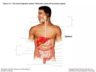
Stomach Anatomy • Located on the left side of the abdominal cavity • C-shaped • Nearly hidden by the liver and diaphragm
Stomach Anatomy • Regions of the stomach • Cardiac region – near the heart • Fundus – expanded part lateral to the cardiac region • Body - midportion • Pylorus – funnel-shaped terminal end
Stomach Anatomy • Food enters the stomach at the cardioesophageal sphincter • Food empties into the small intestine at the pyloric sphincter
Stomach Anatomy • The stomach is about 10 inches long • Its diameter depends on how much food it contains • When it is full it can hold about 1 gallon of food • When it is empty it collapses inward on itself
Stomach Anatomy • Rugae – internal folds of the mucosa (seen especially when the stomach is empty) • External regions • Lesser curvature – the convex lateral surface of the stomach (smaller curve) • Greater curvature – the concave medial surface of the stomach (larger curve)
Stomach Anatomy • There are two layers of peritoneum that are attached to the stomach: • Lesser omentum – attaches the liver to the lesser curvature • Greater omentum – attaches the greater curvature to the posterior body wall • Drapes downward and covers the abdominal organs like a lacy apron • Contains fat which helps to insulate, cushion, and protect abdominal organs
Peritonitis • Peritonitis – occurs when the peritoneum is infected • The peritoneal membranes tend to stick together around the infection site • Helps to seal off and localize many intraperitoneal infections • Provides time for macrophages to mount an attack
Stomach Functions Acts as a storage tank for food Site of food breakdown Chemical breakdown of protein begins After food has been processed in the stomach, it delivers chyme (processed food) to the small intestine
Structure of the Stomach Mucosae • Besides the usual longitudinal and circular muscle layers, the stomach contains a third obliquely arranged layer in the muscularis externa. • Does more than just move food along the tract • Allows it to churn, mix and pummel the food, physically breaking it down to smaller fragments
Specialized Mucosa of the Stomach • The mucosae of the stomach is a simple columnar epithelium that produces large amounts of mucus. • It dotted with deep gastric pits (formed by folded mucosa) • Glands and specialized cells are in the gastric gland region
Specialized Mucosa of the Stomach • Mucous neck cells – produce a sticky alkaline mucus • Clings to the stomach mucosa and protects the stomach wall itself from being damaged by the acid and digested by the enzymes • Gastric glands – secrete gastric juice
Specialized Mucosa of the Stomach Chief cells – produce protein-digesting enzymes (pepsinogens) Parietal cells – produce hydrochloric acid, which makes the stomach acidic and activates the enzymes Endocrine cells – produce gastrin (important for digestion)
Small Intestine • The body’s major digestive organ • Site of nutrient absorption into the blood • Muscular tube extending from the pyloric sphincter to the ileocecal valve • Longest section of the alimentary canal (6-13 feet in a living person) • Is encircled and framed by the large intestine
Small Intestine: Three Subdivisions • Duodenum • Attached to the stomach • Curves around the head of the pancreas • About 10 inches long • Jejunum • Extends from the duodenum to the ileum • About 8 feet long • Ileum • Terminal part of the small intestines • About 12 feet long
Chemical Digestion in the Small Intestine • The small intestine is able to process only a small amount of food at one time • The pyloric sphincter (literally, “gatekeeper”): • Controls food movement into the small intestine from the stomach • Prevents the small intestine from being overwhelmed
Chemical Digestion in the Small Intestine • Enzymes complete the chemical breakdown of food in the small intestine • Source of enzymes that are mixed with chyme • Produced by the intestinal cells • Produced by the pancreas and ducted into the duodenum of the small intestine via the pancreatic duct
Chemical Digestion in the Small Intestine • Bile enters the duodenum from the gall bladder via the bile duct • Bile is formed by the liver • Bile helps to break down fats
Main Function of the Small Intestine • Nearly all food absorption occurs in the small intestine • Its wall has three structures that increase the absorptive surface tremondously • Microvilli • Villi • Circular Folds • Well suited for absorption • These structural modifications decrease in number toward the end of the small intestine
Villi of the Small Intestine • Villi - Fingerlike structures of the mucosa • Give the small intestine more surface area • Gives it a velvety appearance
Microvilli of the Small Intestine • Microvilli - small projections of the plasma membrane • Gives the cell surface a fuzzy appearance • Sometimes referred to as brush border • Found on absorptive cells
Structures Involved in Absorption of Nutrients • Digested foodstuffs are absorbed through the villus of mucosa cells via: • Rich capillary bed • A modified (specialized) lymphatic capillary called a lacteal
Folds of the Small Intestine • Circular folds, called plicae circulares, are deep folds of both the mucosa and submucosa layers • Do not disappear when filled with food (unlike the rugae)
Folds of the Small Intestine • The submucosa has Peyer’s patches (collections of lymphatic tissue) • More are found towards the end of the small intestine • The undigested food residue in the intestine contains huge numbers of bacteria, which must be prevented from entering the bloodstream