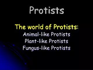
Exploring Protists: Classification and Lab Studies in Eukaryotes
This lab session delves into the fascinating world of protists within the eukaryotic domain, known for their diversity and complexity. As we explore various classifications, we will examine Euglenozoans, Alveolates, and Stramenopiles, utilizing prepared slides and wet mounts to observe organisms like Euglena, Paramecium, and diatoms. Students will engage in hands-on microscopy to study their morphology, ecological roles, and economic significance. This exploration will spark questions about these organisms and their importance in our ecosystems.

Exploring Protists: Classification and Lab Studies in Eukaryotes
E N D
Presentation Transcript
Protists I Lab 3 BIOL 171
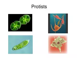
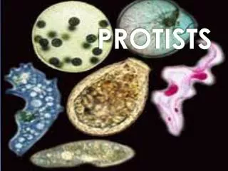
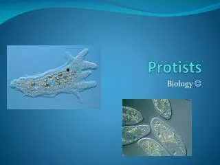
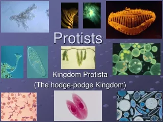
We’ll be looking at all of these! Protists are everywhere in Eukarya! “the junk drawer of the eukaryotes” Ancestral Eukaryote
We’ll be looking at all of these! Protists are everywhere in Eukarya! “the junk drawer of the eukaryotes” Ancestral Eukaryote
6 Kingdoms • Plants (Plantae) • Animals (Animalia) • Fungi (Fungi) • Eubacteria • Archaeabacteria • Protista
Lab Study A: Euglenozoans • Trypanosomalevisi(prepared slide) • Euglena (make wet mount) – not in manual (use depression slide) • Termites (Trichonympha) - procedure not in manual
Trichonympha • Lives in the intestine of the termite • Bacterial endosymbionts inside Trichonympha digest cellulose - Termite > Trichonympha > Spirochetes Procedure • Place a couple of drops of Ringer’s solution on a clean slide. • Transfer a termite into the drop of solution. • Place slide under a dissecting microscope. • Place the tips of dissecting needles at either end of the termite and pull in opposite directions. • Locate the long tube that is the termite’s intestine. • Place a cover slip over the specimen and lightly press down on coverslip to release the Trichonympha from the intestines. Observe with a compound microscope.
Lab Study B: Alveolates Ciliate: Paramecium caudatum – (wet mount) in manual Dinoflagellates: mixed dinoflagellates (live & wet mount), and Peridinium (wet mount) not in manual
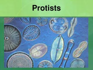
Lab Study C: Stramenopiles Diatoms (Bacillariophyta) – make wet mount Also observe diatomaceous earth (the cell wall deposits from diatoms) – make wet mount and look at prepared slides
Brown Algae (Phaeophyta) Living: Ectocarpus and Sphacelaria Preserved: Fucus and Laminaria
Lab Study D: Rhizaria(different title from manual) • Foraminiferans - prepared slides
Think about… • Morphological characteristics • Ecology of the organism • How does the organism get around? • What role do they play in the ecosystem? • Do they have any economic value? • Where do they live? • Don’t know the answer?? It’s probably a great research question! Ask me about it.