Download
1 / 17
170 likes | 471 Views
DVT: Symptoms and work-up. Sean Stoneking. DVT Epidemilogy. Approximately 600,0000 new cases of DVT each year 50% in hospitalized patients or nursing home residents. Clinical Signs and Symptoms. Up to 50% are asymptomatic Pain Edema Warmth Discoloration

E N D
DVT: Symptoms and work-up Sean Stoneking
DVT Epidemilogy • Approximately 600,0000 new cases of DVT each year • 50% in hospitalized patients or nursing home residents
Clinical Signs and Symptoms • Up to 50% are asymptomatic • Pain • Edema • Warmth • Discoloration • Palpable cord of a thrombosed vein • Homan’s sign (present 1/3 of cases)
DDx of acute edema/leg pain? • Infection • Trauma/injury • Venous insufficiency
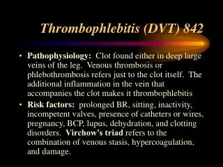
Risk Factors: Virchow’s Triad • Stasis • Venous endothelial injury • Hypercoagulable state
Risk factors • Past DVT • Immobilization • Pregnancy • OCP and HRT • Trauma • Obesity • Age • Sepsis • Cancer • Diseases that alter blood viscosity (sickle cell, polycythemia, multiple myeloma)
Risk Factors: Thrombophilias • Anticoagulant protein deficiency (Protein C/S, Antithrombin Plasminogen, Heparin cofactor II) • Dysfibrinogenemia • Antiphospholipid antibodies • Factor V Leiden mutation (heterozygous) • Prothrombin G20210A mutation (heterozygous)
Clinical features 1. Active cancer (treatment within 6 months) 2. Paralysis, paresis, or immobilization of lower extremity 3. Bedridden for more than 3 days because of surgery (within 4 weeks) 4. Localized tenderness along distribution of deep veins 5. Entire leg swollen 6. Unilateral calf swelling of greater than 3 cm (below tibial tuberosity) 7. Unilateral pitting edema 8. Collateral superficial veins 9. Alternative diagnosis as likely as or more likely than DVT Points 1 1 1 1 1 1 1 1 -2 Wells pretest probability
Pretest Probability Interpretation • >=3 points: high risk (75%) • 1 to 2 points: moderate risk (17%) • <1 point: low risk (3%).
Testing Modalities • Ulrasonography • D-dimers • Contrast venography • MRI • Spiral CT
Ultrasound • In the proximalveins sensitivity is 97% • In the calf veins sensitivity is only 73% • It cannot distinguish between an old clot and a new clot.
D-dimers • Degradation product of a cross-linked fibrin bloodclot. • Levels also elevated in recent major surgery,hemorrhage, trauma, pregnancy or cancer. • Sensitive but nonspecific • The value is in a negative test result
Contrast venography • “Gold Standard” for imaging DVT • can image entire lower extremities • Sensitive in asymptomatic patients • Limitations: invasive, contrast reactions
MRI • Good test for suspected iliac vein or inferior vena caval thrombosis • More sensitive than any other noninvasive study in suspected calf vein thrombosis. • Expense, lack of general availability
Spiral CT • When PE is suspected, can scan chest and lower extremities with same amount of contrast. • ~50% more costly than ultrasound • Risk of contrast reaction
Summary • Use risk stratification of H&P with diagnostic testing to make the diagnosis • Up to 50% of pts with DVT are asymptomatic • Negative D-dimer rules out DVT in low probability • Ultrasound useful for diagnosing proximal thromboses • MRI and contrast venography useful for diganosing distal thromboses