Understanding Risk Factors for Venous Thromboembolism (VTE) and Deep Vein Thrombosis (DVT)
This comprehensive overview outlines the various risk factors associated with Venous Thromboembolism (VTE) and Deep Vein Thrombosis (DVT). Key elements include acquired factors like age, cancer, and immobilization, as well as inherited thrombophilia. The document explores significant statistics, including which cancers are most likely to result in VTE. Additionally, it details the Wells Score as a diagnostic tool for DVT, highlighting its predictive value based on pretest probability. Understanding these factors is vital for prevention and effective management of VTE and DVT.


Understanding Risk Factors for Venous Thromboembolism (VTE) and Deep Vein Thrombosis (DVT)
E N D
Presentation Transcript
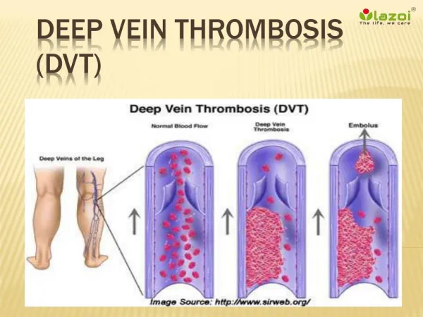
DVT Santosh Reddy,MD FACP
Risk factors for VTE • Age >75 years • Cancer • History of prior VTE • Virchow’s triad • Alteration in blood flow • Injury to endothelium • Thrombophilia
ACQUIRED DISORDERS Malignancy Presence of a central venous catheter Surgery, especially orthopedic Trauma Pregnancy Oral contraceptives INHERETED THROMBOPHILIA Factor V Leiden mutation Prothrombin gene mutation Protein S deficiency Protein C deficiency Antithrombin (AT) deficiency Rare disorders Dysfibrinogenemia Causes for DVT
Acquired Risk • Hormone replacement therapy • Tamoxifen • Immobilization • Congestive failure • Antiphospholipid antibody syndrome • Myeloproliferative disorders • Polycythemia vera • Essential thrombocythemia • Paroxysmal nocturnal hemoglobinuria • Inflammatory bowel disease • Nephrotic syndrome • Hyperviscosity • Waldenstrom's macroglobulinemia • Multiple myeloma • Marked leukocytosis in acute leukemia • Sickle cell anemia
Which cancers are most likely to give you VTE • A Danish study evaluated almost 27,000 patients with VTE • Lung — 17 percent • Pancreas — 10 percent • Colon and rectum — 8 percent • Kidney — 8 percent • Prostate — 7 percent
Physical Findings sensitivity, specificity, and accuracy for DVT. • Edema — 97, 33, and 70 percent • Pain — 86, 19, and 58 percent • Warmth — 72, 48, and 62 percent • Homan’s sign is unreliable (an increased resistance to dorsiflexion of the foot ,associated discomfort or pain)
Differential for DVT • Muscle strain, tear, or twisting injury to the leg • Leg swelling in a paralyzed limb • Lymphangitis or lymph obstruction • Venous insufficiency • Popliteal (Baker's) cyst • Cellulitis • Knee abnormality • Unknown
Wells Score, why it helps • One report of 593 patients with suspected DVT validated a measure of pretest probability (Wells score, show table 2 and show calculator). • DVT was documented in 3, 17, and 75 percent of patients with low, moderate, or high pretest probabilities, respectively.
Pretest Wells Score 1 point for any of the below • Active Cancer (tx within the previous 6mos or palliative) • Paralysis, paresis, recent plaster immobilization of LE. • Recently bedridden for more than 3 days or major surgery, within 4 weeks • Localized tenderness along the distribution of the deep venous system • Entire leg swollen • Calf swelling my more than 3 cm when compared to the asymptomatic leg (measured belo tibial tuberosity) • Pitting edema (greater in the sxs leg) • Collateral superficial veins (varicose don’t count) Alternative diagnosis as likely or more likely than that of DVT (-2) (-2) – 0 Points: Low Probability 1 – 2 Points: Moderate Probability 3 – 8 Points: High Probability
Probability DVT • High Probability; 85% DVT 3 major risk factors no alternative Diagnosis • Intermediate ;35 % DVT • Low DVT ;5 % DVT
Spontaneous VTE Paget-Schroetter syndrome • due to an underlying compressive anomaly at the thoracic outlet. • compression of the vein either between the first rib and a hypertrophied scalene or subclavius tendon or between these tendons themselves. • Compression between the clavicle and a cervical rib as well as partial occlusion of the vein by a congenital web have also been reported.
May – Thurner Syndrome May-Thurner syndrome — Hemodynamically significant compression of the left common iliac vein between the overlying right common iliac artery and the underlying vertebral body (May-Thurner syndrome) is a common anatomic pattern in normal subjects, which has been associated with unprovoked left iliofemoral DVT or chronic venous insufficiency. Visualization of a clot this high in the pelvis may be difficult to detect using ultrasonography, and, if DVT is strongly suspected, further testing should be performed using contrast venography, magnetic resonance imaging, or intravascular ultrasound imaging
Inferior Vena Cava Abnormalities • Inferior vena cava abnormalities — Congenital absence, hypoplasia, or malformation of the inferior vena cava (IVC) may lead to DVT formation. • primarily occurs in young patients and may be bilateral • Study in Update shows, in a series of 97 consecutive patients presenting with confirmed DVT of the lower extremities, 5 of the 31 with thrombotic occlusion of the iliac veins had an anomaly of the IVC; thrombosis was bilateral in one and recurrent in two.