Download
1 / 5
50 likes | 81 Views
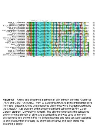
Amino acid sequence alignment of PilA and OxpG from G. sulfurreducens along with pilins and pseudopilins from various bacteria. Alignment optimized through Clustal X and SeAl to create phylogenetic tree. FeOOH, NTA, AQDS concentrations effects on Fe(III) reduction by PilA mutant analyzed. Characterization of G. sulfurreducens and Shewanella Pili using CP-AFM. Pseudomonas aeruginosa strain K pili analysis.

E N D
Figure S1 Amino acid sequence alignment of pilin domain proteins (GSU1496 (PilA) and GSU1776 (OxpG)) from G. sulfurreducens and pilins and pseudopilins from other bacteria. Amino acid sequence alignments were first generated using the Clustal X (1.8) program and manually optimized using the SeAl v. 2.0a11 Carbon program (University of Oxford). The alignment containsthe conserved amino-terminal domain of pilins and pseudopilins and was used to infer the phylogenetic tree shown in Fig. 1c. Different amino acid residues were assigned to one of a number of groups (by chemical similarity) and each group was assigned a colour.
30 FeOOH + NTA +AQDS 25 20 15 Fe(II) (mM) 10 5 0 wild-type PilA- mutant Figure S2 Restoration of Fe(III) oxide (FeOOH) reduction by the PilA- mutant after media supplementation with 4 mM NTA, an Fe(III) chelator, or 0.1 mM AQDS, an electron shuttle that alleviates the need for establishing contact with the insoluble Fe(III) oxides. The levels of soluble Fe(II), produced as a result of the reduction and growth on Fe(III) oxides, were measured after 3 weeks of incubation with the insoluble electron acceptor.
b 0.1 µm c 10 nm a 0.1 µm Figure S3 TEM to negatively stained wild-type pili. (a-b) G. sulfurreducens cells expressing pili after 48h of incubation at 250C in medium with fumarate. (c) Pili filaments that have been mechanically sheared off the outer surface of the wild-type strain of G. sulfurreducens. The wild-type pili preparations were analyzed by conducting-probe AFM (Fig. 4) in order to study the electroconductive properties of G. sulfurreducens pili.
a b 20 nm 1 nA 100 nm c 100 mV Figure S4 CP-AFM analyses to Shewanella oneidensis MR-1 pili. (a) Height image of a pilus filament. (b) Current map obtained while applying a sweeping voltage (c) showing no detectable conductivity across the pilus filament.
a b 15 nm 1 nA 100 nm c 200 mV Figure S5 CP-AFM analyses to Pseudomonas aeruginosa strain K pili. (a) Height image of several pilus filaments. (b) Current map obtained while applying a sweeping voltage (c) showing no detectable conductivity across the pilus filament.


![[Fe II ] 1.64 m m](https://cdn1.slideserve.com/3423313/slide1-dt.jpg)

 2 , X = BF 4 , ClO 4](https://cdn2.slideserve.com/5360123/fe-ii-high-spin-low-spin-transition-in-fe-ii-isoxazole-6-x-2-x-bf-4-clo-4-dt.jpg)