
Nervous system
Nervous system. Nervous system - organization. Somatic NS Receptors Integration Effectors 5 senses Cortex (& assoc.) Skeletal muscles Autonomic NS Receptors Integration Effectors Chemo-baro hypothalamus cardiac, Osmo- medulla oblongata smooth

Nervous system
E N D
Presentation Transcript
Nervous system - organization Somatic NS Receptors Integration Effectors 5 senses Cortex (& assoc.) Skeletal muscles Autonomic NS Receptors Integration Effectors Chemo-baro hypothalamus cardiac, Osmo- medulla oblongata smooth Receptors muscles glands
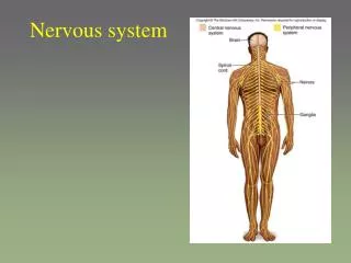
Nervous system - Outline • Nervous tissue • Central Nervous System = CNS • Brain • Spinal cord • Peripheral Nervous System = PNS • Cranial nerves • Spinal nerves • Autonomic Nervous System (a special case)
Nervous system - Outline • Nervous tissue • Central Nervous System = CNS • Brain • Spinal cord • Peripheral Nervous System = PNS • Cranial nerves • Spinal nerves • Autonomic Nervous System (a special case)
Nervous tissue • Neuron – main cell • Neuroglia – supporting cells • Astrocytes • Microglia • Oligodendrocytes • Schwann cells • Ependymal cells
Neurons • Neurons are classified by their shape • Multipolar = 1 axon + multiple dendrites (CNS) • Bipolar = 1 axon, 1 dendrite (senses) • Unipolar = “T”-shaped neuron (ganglia) • Axomenic (anaxonic) = multiple dendrites…NO axons!!! NO action potentials (adrenal gld) • Based on function: • Sensory: conduct nerve impulse to the brain • Motor: conduct nerve impulse to the periphery. From the brain
90% of nervous tissue cells Do not have transmission properties Provide structural support and protect the integrity of the nervous tissue 5 types of cells: - Astrocytes - Microglia - Ependyma cells - Oligodendrocytes - Schwann cells Neuroglia
Provide physical support for neurons Control movements of nutrients and wastes to and from neurons Help recycle and process some neurotransmitters Play a role in the Blood-Brain Barrier (BBB) Play a role in synapse formation Maintain constant brain ECF Astrocytes
Protects the brain from “external” influences Inability of some compounds to cross from the blood into the brain Due to tight junctions between endocytes of he capillary wall Maybe due to astrocytes Glucose, gases can pass Some medications and other compounds can not pass Blood-brain barrier
Derived from macrophages Defense of the brain Clean up old, sick and dead cells Microglia
Line the ventricles of the brain and form, with the blood vessels, the choroid plexuses Secrete the cerebrospinal fluid (CSF) Ependymal cells
Found in the peripheral nervous system (PNS) only Form myelinated sheath Schwann cells
Present in the white matter of the CNS only One cell sends cytoplasmic extensions containing myelin toward several neurons Each extension wraps around a portion of the neuronal axon Oligodendrocytes
Myelin • The myelin sheath acts as an insulating layer • Remember that in the CNS, myelin is formed by oligodendrites, and in the PNS, myelin = Schwann cells • Myelination (the wrapping of neurons) starts during fetal development, BUT, does not complete until adolescence • Low fat diets for children contraindicated • Myelin is high in phospholipids
Myelin • Why myelin? • Allows for a fast response (10X faster than unmyelinated axon) • Usually reserved for nerve fibers that innervate organs/tissues that are important for speed • Not all neurons will be myelinated • If every neuron were myelinated, the neural mass would be far too big (myelin adds mass to the neuron) • “slow” unmyelinated fibers are fine for areas where you don’t really need an instant response • Dilating pupils, secreting stomach acid etc.
Ensheathed or “unmyelinated neurons” The classic “myelinated neuron” vs. the “confusing” ensheathed neuron. Un-myelinated neurons are not truly “bald”, but are ensheathed in a Schwann cell as shown to the right
http://www.pathology.vcu.edu/WirSelfInst/cerebrovasc.html During a cerebral infarction (blood clot leading to lack of blood flow to a region of the brain), neurons will die. They will be replaced by astrocytes that form a dense, almost calcified structure. The process through which astrocytes form this scar tissue = astrocytosis. The end result is a cyst in the site of infarction.
Multiple Sclerosis (MS) • Autoimmune disease that attacks the myelin sheat • Damaged myelin = reduced axon conductivity • Often occurs in “attacks”…episodes of autoimmune attacks • In early years = slower to fully develop • In elderly, development is much more rapid • No real effective treatment
Neuron function: Action potentials • Neurons communicate between themselves and make decision through action potentials ( wave of depolarization sweeping down the axon) and signal transduction at the synapse • You will learn the mechanism in human physiology
Nervous system - Outline • Nervous tissue • Central Nervous System = CNS • Brain • Spinal cord • Peripheral Nervous System = PNS • Cranial nerves • Spinal nerves • Autonomic Nervous System (a special case)
Brain and spine development • During fetal development: • Band of tissue called “neuroectoderm” appears along the back and develops into the “neural plate • Neural plate wraps on itself, forming neural groove, and eventually, the neural tube • The process is dependant on adequate the mother’s folate levels • Neural tube eventually develops 3 “bulges” • http://learningobjects.wesleyan.edu/neurulation/animation.php
Brain and spinal cord developments: abnormalities • During fetal development, failure of the posterior tube to close: • Spina bifida (neural tube development, all impacted by folic acid level) • Anencephaly (failure of anterior part of neural tube to close)
The brain • General considerations • Meninges • Cerebrospinal fluid • Brain matter • Forebrain • Cerebrum • Diencephalon • Limbic system • Brainstem • Midbrain • Pons • Medulla oblongata • Cerebellum • Reticular formation
The brain • General considerations • Meninges • Cerebrospinal fluid • Brain matter • Forebrain • Cerebrum • Diencephalon • Limbic system • Brainstem • Midbrain • Pons • Medulla oblongata • Cerebellum • Reticular formation
Brain - Meninges Like in the spinal cord: 3 layers Dura mater Arachnoid Pia mater Spaces between: Cranium-dura mater = no space Dura mater-arachnoid = subdural space -> connective tissues, blood sinuses Arachnoid-pia mater = subarachnoid space CSF Pia mater – brain = no space
The brain • General considerations • Meninges • Cerebrospinal fluid • Brain matter • Forebrain • Cerebrum • Diencephalon • Limbic system • Brainstem • Midbrain • Pons • Medulla oblongata • Cerebellum • Reticular formation
Brain – Ventricles and CSF • Ventricles: chambers within the brain • 4 ventricles: • 2 lateral • “third ventricle” • “fourth ventricle” • Ventricles are connected: -Interventricular foramen connect the 2 lateral ventricles and third ventricle • Cerebral aqueduct connect third and fourth ventricles • The lateral apertures allow circulation of the CSF into the subarachnoid space • The median aperture connect fourth ventricle to the central canal, into the spinal cord
Cerebrospinal fluid - CSF • Three main functions: • Maintains brain at neutral buoyancy (brain matter is too soft) • Protects the brain from striking the cranium • Chemical stability • CSF acts to wash out the metabolic wastes of the CNS into the blood supply
CSF • Secreted by the ependymal cells lining the ventricles. With connective tissue and blood vessels, they form the choroid plexus • Secreted CSF travels through the ventricle and exits into the subarachnoid space. • CSF is reabsorbed by the arachnoid granulations, located in the arachnoid meninges
The brain • General considerations • Meninges • Cerebrospinal fluid • Brain matter • Forebrain • Cerebrum • Diencephalon • Limbic system • Brainstem • Midbrain • Pons • Medulla oblongata • Cerebellum • Reticular formation
Brain – Grey and white matter • Grey matter : • body of neurons + unmyelinated axons • Surface cortex • Deep nuclei (clump of grey matter) • White matter • within the grey matter, made of bundles of nerve fibers/tracts relaying information from 1 part of the brain to another
Brain – Grey matter • Integration of sensory input and output • Cerebral cortex (surface of the brain) • 6 layers in human form the neocortex: I, II, III, IV, V, and VI • In nuclei
Brain – white matter Bundled into tracts • Projection tracts: from the brain stem to the cerebrum (vertical lines of traffic) • Commissural tracts: from 1 hemisphere to the other, usually via the corpus callosum • Horizontal lines of traffic • Association tracts: within the same hemisphere, connecting different regions of the cerebrum • “internal” lines of traffic
The brain • General considerations • Meninges • Cerebrospinal fluid • Brain matter • Forebrain • Cerebrum • Diencephalon • Limbic system • Brainstem • Midbrain • Pons • Medulla oblongata • Cerebellum • Reticular formation
Forebrain – Cerebrum: general view Frontal lobe: voluntary motor control, motivation, foresight, memory, mood, aggression/social judgment Parietal lobe: sensory reception and integration Occipital lobe: vision Insula: understanding spoken language, taste, sensory integration from visceral organs Temporal lobe: hearing, smell, memory, visual recognition, emotions
Cerebrum • General considerations • The lobes • Frontal lobes • Parietal lobes • Temporal lobes • Occipital lobes • Basal ganglia
Cerebrum • General considerations • The lobes • Frontal lobes • Parietal lobes • Temporal lobes • Occipital lobes • Basal ganglia
Brain anatomy • cerebrum • Right and left sides (hemispheres), separated by “longitudinal fissure” • They’re not truly separate; deep to the fissure is a connection between both sides = corpus callosum • Other landmarks: • Central sulcus divides the frontal lobe of the cerebrum from the parietal lobe • Most dorsal (caudal), right above the cerebellum = occipital lobe
Cerebrum • General considerations • The lobes • Frontal lobes • Parietal lobes • Temporal lobes • Occipital lobes • Basal ganglia
The cerebral hemispheres • Composed of 5 types of lobes and internal nuclei • Frontal • Parietal • Temporal • Insula • Occipital • Internal nuclei:
Frontal lobe Many functions: - personality - Problem solving - motor regions of the brain • In fact, they’re adjacent to the respective sensory areas, on the pre-central gyrus • Thus, you can also create a motor humunculus • Signals from these regions travel to the brain stem • Decussate in the neck and onward to their target muscle groups • The basal nuclei are important for coordinating motor function • Feedback signals of muscle movements (from the various muscle spindle formations)
Frontal lobe: motor area • The lower region, where the mouth is located, is the Broca’s area (on the left side only, in 90% of people) Broca’s area controls speech
Parietal lobes • Receive sensations from the body (primary sensory area) • Interpret sensations (interpretative or associated sensory area)
Temporal lobes • Primary auditory area: receive auditory impulses from the ear • Associated auditory area: interpret auditory impulses • Wernicke’s area (partially in parietal); make senses of the auditory, visual and touch info • Hippocampus: involved in memory • Amygdala is important for feelings, especially rage
Occipital lobes • Primary visual area: receives impulses from retina • Associated visual area: interpret visual impulses
Cerebrum • General considerations • The lobes • Frontal lobes • Parietal lobes • Temporal lobes • Occipital lobes • Basal ganglia
Basal nuclei • Buried deep into the brain matter • There are at least 3 “nuclei” or neural control centers • These nuclei are involved in relaying neural signals from the substantia nigra region of the midbrain (on the brainstem, but a separate region of the brain) • Also relay signals from the motor centers of the cerebral cortex • Help control smoothness of movement, muscle tension and posture
The basal nuclei consists of at least 3 regions of the deep cerebrum: Caudate nucleus, Putamen and Globus palidus (all 3 together are known as the corpus striatum)


