Download

1 / 57
570 likes | 964 Views
CH R ONIC. SUPPUR A TIVE. O TITIS. MEDIA. (CSOM). OBJ E C TIVE S. W h a t is C S OM T ype of CS O M E t i ology P a tho l ogy F e a tu r e I n v e s t i g a t i on T r e a tme n t. INT R ODU C TION Lon g - st and i ng i n f ect i on of a part or

E N D
CHRONIC SUPPURATIVE OTITIS MEDIA (CSOM)
OBJECTIVES What is CSOM Type of CSOM Etiology Pathology Feature Investigation Treatment
INTRODUCTION Long-standinginfectionof a partor the middleear cleftcharacterized –Byeardischarge –Apermanentperforation. • whole of
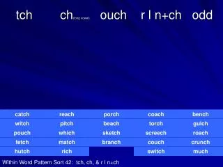
TYPESOFCSOM Tubotympanic.Alsocalledthesafeorbenigntype; Involvesanteroinferiorpartofmiddleearcleft, i.E.Eustachiantube andmesotympanum Associatedwithacentralperforation. Thereisnoriskofseriouscomplications. 1. • it • • 2. • Atticoantral.Also calledunsafeordangerous Involvesposterosuperiorpartofthecleft (I.E.Attic,antrumandmastoid) type; it • • Associatedwithanatticoramarginalperforation. The diseaseis oftenassociatedwith abone eroding process suchascholesteatoma,granulationsorosteitis. Riskofcomplicationsishighinthisvariety. •
Tubotympani (safe) Atticoantral(unsafe) Discharge Profuse,mucoid, odourless Central Uncommon Pale Absent Rare Scanty,purulent, smelling foul Perforation Granulations Polyp Cholesteatoma Complications Audiogram Attic ormarginal Common Redandfleshy Present Common Mildto moderate Conductive deafness ormixed conductive deafness
ETIOLOGY • • Thedisease startsinchildhood Itis thesequela of acuteotitis. –Theperforationcentraland becomes permanentand permitsrepeatedinfectionfromtheexternalear. Ascending infectionsviatheeustachiantube. –Infectionfrom tonsils,adenoidsandinfectedsinuses may beresponsibleforpersistentor recurring otorrhoea. Persistent mucoidotorrhoea is sometimesthe result of allergytoingestantssuch as milk, eggs, fish, etc. • •
PATHOLOGY 1. 2. Perforationof pars tensa. Middle ear mucosa. Itis oedematousand velvety when disease Polyp. is active. 3. Itis usuallypalein contrasttopink,fleshypolyp seen in atticoantraldisease Ossicular chain. Itis usuallyintactand mobilebut mayshow some degree necrosis,particularlyofthe longprocessofincus. Tympanosclerosis. 4. of 5. Itis seen aswhitechalkydeposit on thepromontory,ossicles, joints, tendons and ovaland roundwindows and interfere with the mobilityof thesestructuresand cause conductive deafness. Fibrosisand adhesions. 6.
BACTERIOLOGY • Pus culture inbothtypes of aerobic anaerobic CSOM Commonaerobic organisms and • – – – – Pseudomonasaeruginosa, Proteus, Escherichiacoli Staphylococcusaureus, • AnaerobesincludeBacteroides anaerobic Streptococci. fragilisand
CHRONIC OTITISMEDIA Mucosal Squamosal diseases diseases Active Inactive Healed Active Retract Retract pockets Retraction
CLINICALFEATURES 1. Eardischarge. It isnonoffensive,mucoidormucopurulent, intermittent. 2. Hearing loss. constant or Itis conductivetype;rarely exceeds (roundwindowshielding effect) 3. Perforation. Alwayscentral 4. Middleearmucosa. 50 dB. Itis seenwhen theperforation is large.
INVESTIGATIONS 1. 2. 3. 4. Examinationundermicroscope Audiogram. Culture andsensitivityof ear discharge. MastoidX-rays/CTscantemporalbone.
TREATMENT • • Auraltoilet. Eardrops. – Antibiotic ear drops polymyxin,chloromycetin,ciprofloxacin are used. – Theyarecombinedwith steroidswhich anti-inflammatory effect. have local • • • • • Systemic antibiotics Precautions Treatmentof contributory Surgicaltreatment Reconstructivesurgery causes
INTRODUCTION Itinvolvespostero-superiorpartof middleear cleft Associatedwith cholesteatoma(bone eroding properties) unsafeordangeroustype. • • •
ETIOLOGY Etiology ofatticoantral disease issame as of cholesteatomaandhas been discussedearlier. Itisseen in scleroticmastoid. • •
PATHOLOGY 1. 2. 3. 4. Cholesteatoma. Osteitisandgranulationtissue. Ossicularnecrosis(cholesteatoma Cholesterolgranuloma. hearer)
BACTERIOLOGY • Same as intubo-tympanictype.
SYMPTOMS 1. Ear discharge. Usually scanty,but alwaysfoul-smelling destruction Hearing loss. dueto bone 2. Hearing isnormalwhenossicularchainis intactor whencholesteatoma,having destroyedtheossicles, bridges the gap causedby destroyedossicles (cholesteatomahearer). 3. Bleeding. Fromgranulationsor the polyp whencleaning theear.
SIGN 1. Perforation. Itis eitheratticorpostero-superior type. 2. Retractionpocket. marginal An invagination of tympanic membrane isseen the atticor posterosuperiorarea ofpars tensa. There are fourstagesoftympanicmembrane retraction. 3. Cholesteatoma. in
StageI •Tympanicmembrane is retractedbut does not contact theincus. StageII •Tympanicmembrane is retracteddeep and contactsthe incus. •Middleear mucosa is not affected.
StageIII •Also calledmiddleear atelectasis. onthe promontoryandossicles. •Middle earspaceis totallyor partiallyobliteratedbut middleear mucosais intact. •Tympanic membrane comes to lie StageIV •Also calledadhesiveotitismedia. and wrapsthe promontoryand ossicles. •Thereis nomiddleear space •Mucosal liningofthe middle ear is absent and tympanicmembrane getsadherent tothepromontory. •Tympanic membrane isvery thin
INVESTIGATIONS 1. 2. 3. 4. Examinationundermicroscope Tuningfork testsandaudiogram. X-raymastoids/CTscantemporal bone. Culture andsensitivityof ear discharge.
FEATURESINDICATING COMPLICATIONS IN CSOM 1. 2. 3. 4. 5. Pain Vertigo Persistentheadache. Facialweaknessindicateserosionoffacialcanal. A listless childrefusingtotake feeds and easily going to sleep(extraduralabscess). 6. 7. 8. 9. Fever,nauseaandvomiting(intracranialinfection). Irritability andneckrigidity(meningitis). Diplopia (Gradenigosyndrome) petrositis. Ataxia(labyrinthitisorcerebellarabscess). 10.Abscessroundtheear(mastoiditis).
TREATMENT 1.Surgical (a)Canalwalldown procedures (b)Canalwallup procedures. 2.Reconstructivesurgery.Hearing can be restoredbymyringoplastyortympanoplasty. 3. Conservativetreatment.
What is cholesteatoma Origin Classification • • •
INTRODUCTION Normally, middle earcleftis lined by differenttypesof epitheliumindifferentregions Itis thepresenceofkeratinizingsquamousepitheliumin themiddle earor mastoid. “Skin in the wrong place.” Essentially,cholesteatomaconsistsof two parts: • • • • (i) the matrix,which is made up ofkeratinizingsquamous epitheliumresting on athin stromaof fibrous tissuesand (ii) a central whitemass, consistingofkeratin debris produced by thematrix . • Also beennamedepidermosisorkeratoma.
ORIGIN 1.Presenceofcongenital cell rests. 2.Invaginationof tympanicmembrane from the attic or posterosuperior partof parstensa in the form ofretractionpockets(Wittmaack’s theory). 3. 4. 5. Basalcell hyperplasia(Ruedi’stheory). Epithelialinvasion(Habermann’stheory). Metaplasia(Sade’stheory).
CLASSIFICATION OFCHOLESTEATOMA • The cholesteatoma is classified into: 1. 2. 3. Congenital Acquired,primary Acquired,secondary
1. CONGENITALCHOLESTEATOMA. • • Itarises fromthe embryonicepidermal 3 importantsites: cell rests – – – Middle ear Petrousapex And thecerebellopontineangle • Presentsas a whitemassbehindanintacttympanic membraneandcausesconductivehearingloss. Itmay sometimesbediscoveredon routineexamination of childrenor atthetimeofmyringotomy. Itmay alsospontaneouslyrupture Presentwitha dischargingearindistinguishablefroma caseofchronic suppurativeotitis media. • • •
2.PRIMARY ACQUIRED CHOLESTEATOMA • No historyof previousotitismedia existingperforation. Theories on itsgenesis are: or a pre- • (A) (B) (C) invaginationofparsflaccida. basalcellhyperplasia. squamousmetaplasia.
3.SECONDARYACQUIRED CHOLESTEATOMA Already apre-existing perforation inpars tensa. Thisis oftenassociatedwithposterosuperior • • marginalperforationor sometimes central perforation. Theories on itsgenesis (A)migrationofsquamousepithelium. (B)Keratinizingsquamousmetaplasia large •
EXPANSIONOF CHOLESTEATOMAAND DESTRUCTIONOFBONE Itinvades the surroundingstructures, firstthe path ofleastresistance,and then byenzymaticbonedestruction. An atticcholesteatomamay extend • • – – – – – – – Backwardsinto theaditus,antrum andmastoid; Downwardsinto themesotympanum; Medially,itmay surroundtheincusand/orhead ofmalleus. Causedestruction ofear ossicles, Erosion ofbonylabyrinth, Canalof facial nerve, Sinus plateortegmentympani • Attributedtovariousenzymessuch as – Collagenase, – Acidphosphatase and proteolytic enzymes, Liberatedbyosteoclastsand mononuclearinflammatorycells, Seeninassociationwithcholesteatoma. • •