Download
1 / 54
570 likes | 836 Views
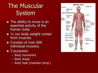
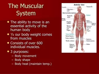
The Muscular System. Muscle Tissue. Made up of muscle fibers (cells) Highly specialized for the active generation of tension Motion by moving skeletal levers Stabilizing body position Regulating organ volume Converts chemical energy to mechanical energy Thermogenesis.

E N D
Muscle Tissue • Made up of muscle fibers (cells) • Highly specialized for the active generation of tension • Motion by moving skeletal levers • Stabilizing body position • Regulating organ volume • Converts chemical energy to mechanical energy • Thermogenesis
Functions of Muscle Tissue • Movement Facilitation • Thermogenesis • Postural Support • Regulation of Organ Volume • Protects Internal Organs • Pumps Blood (HEART)
Characteristics of Muscle Tissue • Contractility • Extensibility (Flexibility) • Elasticity • Excitability (Irritability)
Skeletal Muscle • Attached to bones • Striated appearance under a microscope • Voluntary control (conscious control) • Multinucleated • Sarcolemma - muscle cell membrane • Sarcoplasm - muscle cell cytoplasm • Myofilaments - contractile elements of each muscle fiber
Connective Tissue Components of Skeletal Muscle • Epimysium - surrounds entire muscle • Perimysium - surrounds a bundle of muscle fibers (fascicles) • Endomysium - surrounds each individual muscle fiber • Epimysium, Perimysium, Endomysium converge at the end of a muscle to form a tendon or aponeurosis
Muscle Attachments to Bone • Tendon - round, cord like fibrous connective tissue • Aponeurosis - broad, flat sheet of connective tissue • Tendon Sheaths - specialized tendons that are enclosed tubes of fibrous connective tissue • Contain a film of synovial fluid • Similar in function to bursae
Muscle Tissue Histology • Muscle Fiber - elongated cylindrical cell • Sarcolemma - muscle cell membrane • Sarcoplasm - muscle cell cytoplasm • Myofibrils - groups of contractile myofilaments that run longitudinally within the muscle • Sarcomere - a small section or compartment within a myofibril which serves as the unit of contraction
Muscle Tissue Histology • Myofilaments - structural components of myofibrils • Myosin - thick myofilaments • Actin - thin myofilaments • Z Line - connective tissue that separates individual sarcomeres
Myosin • Thick myofilaments • Occupy the A Band of the sarcomere • Overlap free ends of the actin myofilament • Shaped like a golf club • Long, thick protein molecule (tail) • Globular head at the ends
Actin • Thin myofilaments • Anchored to the Z Line • Two stranded protein molecule intertwined around each other • Associated with two regulatory proteins • Tropomyosin - long stranded protein molecule that follows the contour of actin • Troponin - protein located at regular interval along the tropomyosin that covers the active sites on actin. Has three subunits
Muscle Nerve Interaction • Neuron - nerve cell • Axon - long, threadlike process that transmits impulse away from cell body (may be up to 1 meter in length) • Axon Terminal - branches of the axon after it enters the muscle tissue • Motor Unit - motor neuron and all the muscle fibers it innervates • Neuromuscular Junction - junction between axon terminal and muscle fiber
Muscle Nerve Interaction • Motor End Plate - location on the muscle fiber at the end of an axon terminal • Synaptic End Bulb - distal end of axon terminal • Synaptic Vesicles - membrane enclosed sacs within the synaptic end bulbs that store neurotransmitters
Muscle Nerve Interaction • Synaptic Cleft - space between axon terminal and motor end plate • Subneural Clefts - folds in sarcolemma along the synaptic gutter • Acetylcholine (Ach) - neurotransmitter released from synaptic vesicles that initiates an action potential in a muscle
Sarcoplasmic Reticulum and Transverse Tubules • Sarcoplasmic Reticulum - storage site for calcium within the muscle • Transverse (T) Tubules - invaginations of the sarcolemma deep within the muscle • Terminal Cisternae - dilated end sacs of sarcoplasmic reticulum next to the T-tubules • Triad - a T-tubule and the terminal cisternae on both sides of it
Muscle Action Potential An electrical impulse that originates at the motor end plate, travels along the length of the sarcolemma, down a transverse tubule, and causes the muscle to contract.
Sliding Filament Theory of Muscular Contraction • Due to an action potential, the actin and myosin myofilaments slide past one another shortening the sarcomere • No change in length of myofilaments • H Zone narrows or disappears • I Band narrows or may disappear • A Band remains the same length
Muscle Response to Nervous Stimuli • All or None Principle • Once a threshold stimulus is applied to a motor unit the muscle fibers innervated by that motor unit will contract to their fullest potential • Threshold Stimulus - the weakest stimulus from a neuron that will initiate a muscular contraction
Events Leading to Muscular Contraction • An action potential travels down the motor neuron. When it arrives at the synaptic knob, the membrane of the nerve at the synaptic cleft is depolarized, thereby increasing the Ca++ permeability of the membrane. • Ca++ diffuses from outside of the synaptic knob to inside the synaptic knob.
The influx of Ca++ into the nerve causes the release of Ach. • Ach is ejected into the synaptic cleft, diffuses across the cleft, and depolarizes the muscle membrane. • This increases the permeability of the muscle membrane to Na+. • Na+ rushes into the muscle cell, depolarizing the membrane as it travels away from the motor end plate thus initiating an action potential.
Ach is quickly broken down in the cleft by Ach-ase so that each action potential arriving from the nerve initiates only one action potential within the muscle. • The action potential spreads across the muscle membrane and down the T-tubules deep into the muscle cell. • The action potential of the T-tubules depolarizes the membrane of the nearby sarcoplasmic reticulum which results in the release of Ca++ into the sarcoplasm.
Ca++ is very quickly removed out of the sarcoplasm by the sarcoplasmic reticulum so the effects of one action potential are very short lived and produce a very small contraction. • Many action potentials are necessary to produce enough force to produce a strong or prolonged muscle contraction. • The Ca++ released from the sarcoplasmic reticulum binds with troponin and cause troponin to change shape.
When troponin changes shape, it physically moves the other regulatory protein, tropomyosin, out of the way exposing the active sites on the actin myofilament. • Since the heads or cross-bridges of myosin have a very strong affinity for the active sites on actin, they make contact immediately after the active sites have been exposed. • The acto-myosin complex has ATPase activity and ATP is split into ADP + P and energy is released.
The energy released by the splitting of ATP is used to produce movement of the cross-bridges, sliding the actin and myosin filaments past one another which causes the sarcomere to shorten and the muscle to contract and produce force. • The myosin cross-bridge has a low affinity for ADP but a very high affinity for ATP. • It discards the ADP and becomes recharged with a new ATP.
The myosin then releases its hold on the active sites on actin, swivels back to its original position, and is ready to respond to another action potential. • When another action potential comes along the entire process is repeated. • It takes many action potentials to produce enough shortening of the sarcomeres to generate enough force to produce movement of a body segment.
Cardiac Muscle • Forms the bulk of heart wall (Myocardium) • Striated • Involuntary (typically) • Fibers are quadrangular and branching • Cardiac fibers typically have a centrally located nucleus • Sarcolemmas connected by intercalated discs • Strengthens cardiac muscle tissue • Propagates an action potential from cell to cell through specialized structures on the intercalated discs called gap junctions
Smooth (Visceral) Muscle • Located in walls of hollow internal surfaces such as: • blood vessels - stomach • urinary bladder - intestines • Non-striated in appearance • Involuntary (typically) • Can be stretched to great lengths • Allows for tremendous size variability
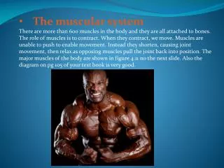
Muscles and Movement • Produce movement by contracting, creating tension on tendons, which pull on bones or other structures. • Most muscles cross over at least one joint. • Attempt to pull bones together. • Since muscles cross over a joint, the joint serves as the axis of rotation and the shortening muscle produces angular rotation.
Muscle Origin and Insertion • Origin • Body segment with most mass • Usually more proximally located • Usually larger surface area of attachment • Insertion • Body segment with least mass • Usually more distally located • Usually smaller surface area of attachment • Gaster (Belly) • Fleshy portion of the muscle between the tendons of the origin and insertion
Roles of Skeletal Muscles • Agonist (Prime Mover) • Muscle responsible for the majority of force • Antagonist • Performs the opposite movement • Synergist • Muscle that assists the agonist • provides additional force • redirects the force of the agonist • Fixator (Stabilizer) • Stabilizes a body segment so the prime mover can act more effectively
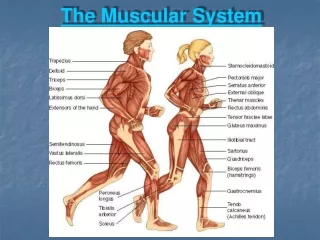
Pectoralis major Deltoid Biceps brachii Sternocleidomastoid Diaphragm Quadriceps rectus femoris vastus medialis vastus lateralis Selected Superficial Skeletal Muscles (Anterior View)
Trapezius Triceps brachii Gastrocnemius Latissimus dorsi Hamstring Group semimembranosus biceps femoris semitendinosus Gluteus maximus Selected Superficial Skeletal Muscles (Posterior View)
Myalgia (Fibromyalgia) • Painful disorders of muscles, tendons, and surrounding soft tissue
Muscular Dystrophies • Muscle destroying diseases characterized by the degeneration of individual muscle fibers • Leads to progressive atrophy of skeletal muscles • Due to a genetic defect