Download
1 / 1
10 likes | 122 Views
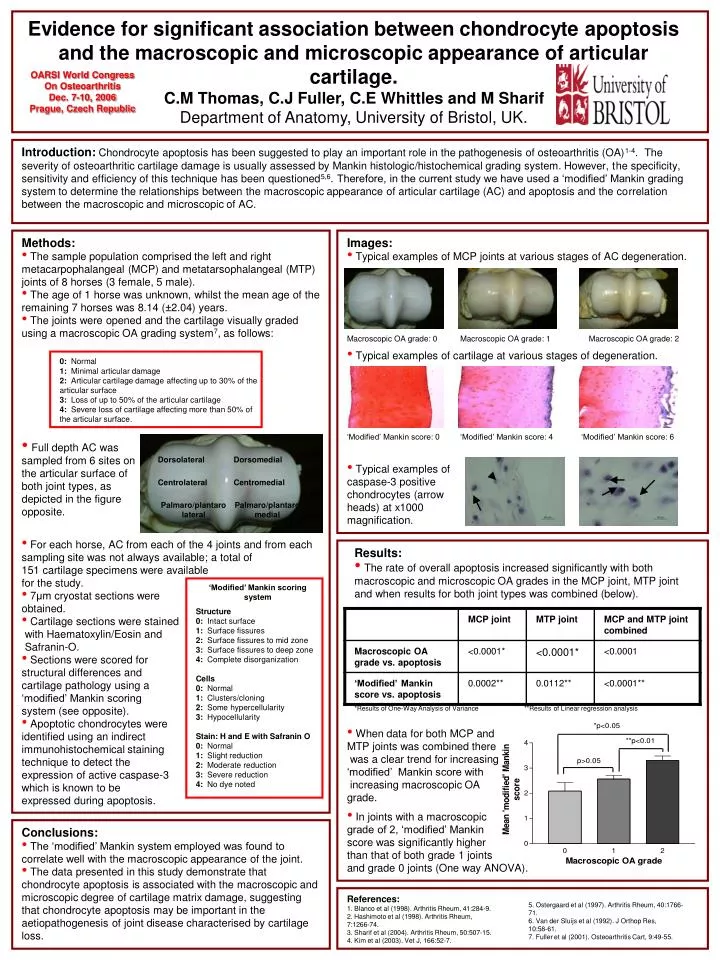
Evidence for significant association between chondrocyte apoptosis and the macroscopic and microscopic appearance of articular cartilage. C.M Thomas, C.J Fuller, C.E Whittles and M Sharif Department of Anatomy, University of Bristol, UK. OARSI World Congress On Osteoarthritis Dec. 7-10, 2006

E N D
Evidence for significant association between chondrocyte apoptosis and the macroscopic and microscopic appearance of articular cartilage. C.M Thomas, C.J Fuller, C.E Whittles and M Sharif Department of Anatomy, University of Bristol, UK. OARSI World Congress On Osteoarthritis Dec. 7-10, 2006 Prague, Czech Republic Introduction:Chondrocyte apoptosis has been suggested to play an important role in the pathogenesis of osteoarthritis (OA)1-4. The severity of osteoarthritic cartilage damage is usually assessed by Mankin histologic/histochemical grading system. However, the specificity, sensitivity and efficiency of this technique has been questioned5,6.Therefore, in the current study we have used a ‘modified’ Mankin grading system to determine the relationships between the macroscopic appearance of articular cartilage (AC) and apoptosis and the correlation between the macroscopic and microscopic of AC. • Methods: • The sample population comprised the left and right metacarpophalangeal (MCP) and metatarsophalangeal (MTP) • joints of 8 horses (3 female, 5 male). • The age of 1 horse was unknown, whilst the mean age of the remaining 7 horses was 8.14 (±2.04) years. • The joints were opened and the cartilage visually graded • using a macroscopic OA grading system7, as follows: • Images: • Typical examples of MCP joints at various stages of AC degeneration. Macroscopic OA grade: 0 Macroscopic OA grade: 1 Macroscopic OA grade: 2 • Typical examples of cartilage at various stages of degeneration. 0: Normal 1: Minimal articular damage 2: Articular cartilage damage affecting up to 30% of the articular surface 3: Loss of up to 50% of the articular cartilage 4: Severe loss of cartilage affecting more than 50% of the articular surface. ‘Modified’ Mankin score: 0 ‘Modified’ Mankin score: 4 ‘Modified’ Mankin score: 6 • Full depth AC was sampled from 6 sites on • the articular surface of • both joint types, as • depicted in the figure opposite. Dorsolateral Dorsomedial • Typical examples of caspase-3 positive chondrocytes (arrow heads) at x1000 magnification. Centrolateral Centromedial Palmaro/plantaro medial Palmaro/plantaro lateral • For each horse, AC from each of the 4 joints and from each sampling site was not always available; a total of • 151 cartilage specimens were available • for the study. • 7μm cryostat sections were • obtained. • Cartilage sections were stained • with Haematoxylin/Eosin and • Safranin-O. • Sections were scored for • structural differences and • cartilage pathology using a • ‘modified’ Mankin scoring • system (see opposite). • Apoptotic chondrocytes were • identified using an indirect • immunohistochemical staining • technique to detect the • expression of active caspase-3 • which is known to be • expressed during apoptosis. • Results: • The rate of overall apoptosis increased significantly with both macroscopic and microscopic OA grades in the MCP joint, MTP joint and when results for both joint types was combined (below). ‘Modified’ Mankin scoring system Structure 0: Intact surface 1: Surface fissures 2: Surface fissures to mid zone 3: Surface fissures to deep zone 4: Complete disorganization Cells 0: Normal 1: Clusters/cloning 2: Some hypercellularity 3: Hypocellularity Stain: H and E with Safranin O 0: Normal 1: Slight reduction 2: Moderate reduction 3: Severe reduction 4: No dye noted *Results of One-Way Analysis of Variance **Results of Linear regression analysis • When data for both MCP and • MTP joints was combined there • was a clear trend for increasing ‘modified’ Mankin score with • increasing macroscopic OA • grade. • In joints with a macroscopic • grade of 2, ‘modified’ Mankin • score was significantly higher • than that of both grade 1 joints • and grade 0 joints (One way ANOVA). • Conclusions: • The ‘modified’ Mankin system employed was found to correlate well with the macroscopic appearance of the joint. • The data presented in this study demonstrate that chondrocyte apoptosis is associated with the macroscopic and microscopic degree of cartilage matrix damage, suggesting that chondrocyte apoptosis may be important in the aetiopathogenesis of joint disease characterised by cartilage loss. References: 1. Blanco et al (1998). Arthritis Rheum, 41:284-9. 2. Hashimoto et al (1998). Arthritis Rheum, 7:1266-74. 3. Sharif et al (2004). Arthritis Rheum, 50:507-15. 4. Kim et al (2003). Vet J, 166:52-7. 5. Ostergaard et al (1997). Arthritis Rheum, 40:1766-71. 6. Van der Sluijs et al (1992). J Orthop Res, 10:58-61. 7. Fuller et al (2001). Osteoarthritis Cart, 9:49-55.