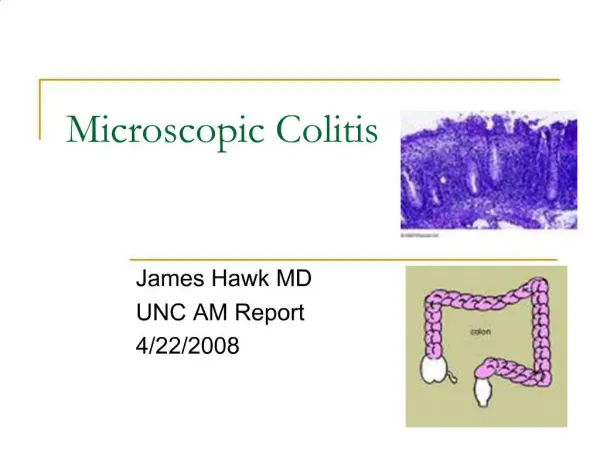
- Microscopic appearance
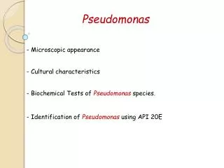
Campylobacter. - Microscopic appearance. - Cultural characteristics. - Biochemical Tests of Campylobacter species. Campylobacters jejuni. Campylobacters coli. Campylobacters are small delicate, spirally curved, motile Gram negative bacteria. Campylobacter jejuni causing:-.


- Microscopic appearance
E N D
Presentation Transcript
Campylobacter - Microscopic appearance - Cultural characteristics - Biochemical Tests of Campylobacter species.
Campylobactersjejuni Campylobacters coli Campylobacters are small delicate, spirally curved, motile Gram negative bacteria
Campylobacter jejunicausing:- - Enteritis. *** Fresh diarrhoeal or dysenteric specimens containing blood, pus and mucus (child under 2 y).
A filtration technique and a non-selective culture medium. A filter of 0.47 μm pore size will retain faecalcommensals and allow Campylobacters to pass through A selective culture medium that contains antimicrobials to inhibit the growth of faecalcommensals.
Blaser’s medium: Containing 10% sheep blood, vancomycin, trimethoprim, polymyxin B, cephalothin, amphotericin B. Skirrow’s blood agar: Containing lyzed horse blood, vancomycin, polymyxin B, trimethoprim Butzlervirion medium: Containing defibrinated sheep blood, cefoperazone, rifampicin, colistin, amphotericin B.
Improved Preston blood free-medium: Containing cefoperazone and amphotericin B. This supplement is added to a Campylobacter blood-free agar base containing bacteriological charcoal, ferrous sulphate, sodium deoxycholate, sodium pyruvate, casein hydrolysate, nutrient broth and agar. Isolations are best on this medium when cultures are incubated at 37 ºC rather than 42–43 ºC C. jejuni produces grey, moist, flat-spreading colonies. Some strains may have a green hue or a dry appearance with or without a metallic sheen.
On Blood agar: C. jejuni and C. coli produce nonhaemolytic spreading, droplet-like colonies
Hippurate hydrolysis: this test can be used to differentiate • C. jejuni from C. coli. • Hippurate is hydrolyzed by C. jejuni and not hydrolyzed by • C. coli.
The end product of hydrolysis of hippuric acid by hippuricase include glycine and benzoic acid. Glycine is deaminated by the oxidizing agent ninhydrin. The end products of the ninhydrin oxidation reacts to form a purple-coloured product.
Helicobacter - Microscopic appearance - Cultural characteristics - Biochemical Tests of Helicobacter species.
- gastric and duodenal ulcers (eradication of H. pylori results in cure and reduces ulcer recurrence in 90% of peptic ulcer patients). - H. pylori also contribute to diarrhoea, malnutrition and growth failure in young children (reduced gastric acid protection leads to infection with enteropathogens). - To isolate H. pylori by culture a gastric biopsy is required. Place a biopsy of mucosa from the gastric antrum in a bottle containing about 0.5 ml of sterile physiological saline. It should reach the laboratory with the minimum of delay.
Using a sterile scalpel and forceps, cut the biopsy into small pieces. Inoculate a plate of chocolate (heated blood) agar or Campylobacter medium, and also place a piece of biopsy in Christensens urea broth
H. pylori appears as a small, spiral or S-shaped Gram negative bacterium
On blood agar, H. pylori colonies are slightly beta-haemolytic. Growth is best at 37 ºC
Urease Test The test organism is cultured in a medium which contains urea and the indicator phenol red. When the strain is urease producing, the enzyme will break down the urea (by hydrolysis) to give ammonia and carbon dioxide. With the release of ammonia, the medium becomes alkaline as shown by a change in colour of the indicator to pink-red.