Download
1 / 19
200 likes | 448 Views
Learn about ion chambers and Geiger-Muller counters for radiation detection. Explore scintillation detectors in gamma cameras, their components, and principles of operation.

E N D
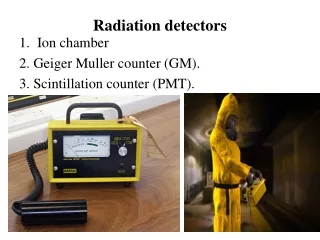
Radiation detectors • Ion chamber 2. Geiger Muller counter (GM). 3. Scintillation counter (PMT).
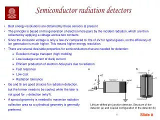
An ion chamber • is a general class of detector that makes use of the electron–ion pairs generated by the passage of radiation through a gas to produce an electrical signal. • It consists basically of two charged plates maintained at different potentials by a voltage supply . • The plates attract electrons or ions, depending on plate polarity, and cause a current pulse i that is proportional to the number of electron–ion pairs produced and to the particle energy if the particle comes to rest in the chamber. • When an ion chamber is used in this way, both to detect the presence of an energetic charged particle and to measure its energy, it is called a proportional counter.
Geiger Muller counter (GM) • The Geiger counter is an instrument used for measuring ionizing radiation used widely in such applications as radiation dosimetry, radiological protection, experimental physics and the nuclear industry. • It detects ionizing radiation such as alpha particles, beta particles and gamma rays using the ionization effect produced in a Geiger–Müller tube; which gives its name to the instrument. • In wide and prominent use as a hand-held radiation survey instrument, it is perhaps one of the world's best-known radiation detection instruments.
Basic components of GM tube. • A Geiger counter consists of a Geiger-Müller tube, the sensing element which detects the radiation, and the processing electronics, which displays the result. • The Geiger-Müller tube is filled with an inert gas such as helium, neon, or argon at low pressure, to which a high voltage typically 400-600 V is applied.
Principle of operation • When a single gamma or beta ray entering the tube, a small amount of ionization is produced. • The center electrode which is at high positive potential attracts the electrons and gives them energy to produce further ionization until the whole volume contains ion pairs. • The electrons are rapidly collected. • The voltage on the center electrode drops and the slow positive ions go to the outer wall. 5. After 400µsec (Dead time) the tube is ready to repeat the Process.
Disadvantages of GM counter • It can not differentiate between the types of ionizing radiation. • It can not differentiate between large and small amounts of ionization which means that it can not measure radiation energy. • It is inefficient for detecting gamma rays. • It can not detect any ionization events in the dead time which means that it is inefficient in detecting high radiation rates (the number of counts will be lower about 104 to 105.
Gamma Camera Scintillation detectors In Nuclear Medicine imaging devices • Is a device used to image gamma radiation radioisotopes this technique is called also scintillation camera. • Gamma camera is used to view and analyze images of the human body or the distribution of the medically ingested, injected or inhaled radionuclides.
Gamma Camera Components 1-Collimators • The collimator provides an interface between the patient and the scintillation crystal by allowing only those photons traveling in an appropriate direction.
Collimators • Types of collimators A) By the accepted energy. B) By the geometric shape. C) By the resolution.
Collimators • By the accepted energy High Energy Collimator Low Energy Collimator Medium Energy Collimator
Collimators • By the geometric shape. Diverging collimator Parallel-Hole Pin-Hole Collimator Converging collimator
Collimators Pin-Hole (more resolution & magnification) hip,thyroid Parallel-Hole Converging لتكبير الصورة وتحديد أفضل للأعضاء Diverging للتصغير في حالة المريض البدين
Detector Scintillation Crystal • The chosen material for the crystal is Na-I (Tl), 40-50 cmdiameter. • The Na-I (Tl) crystal is stationary. • The crystal transform the gamma-ray photon ------> Light photon • Any damage to the crystal results in an inoperable scintillation camera and requires costly replacement of the crystal.
Photomultiplier tube Dynode Connected to High positive volt Photomultiplier tube Photocathode
Photomultiplier tube • The Photocathode transform the light photon --- electron. • The PMT multiplies the electron to be a significant detected signal.
Other circuits • 1)Pre-Amplefier • 2) Amplifier
Advantages of Gamma Camera • The imaging time is only 1-2min. • It can distinguish 2 sources about 5mm apart.