Download
1 / 84
850 likes | 868 Views
Explore the importance of fluid and electrolyte balance for maintaining homeostasis, the role of nurses in preventing and treating disturbances, and the regulation mechanisms of fluid movement. Gain insights into key electrolytes, osmosis, diffusion, and more.

E N D
Fluid and Electrolytes: Balance and DisturbanceDr. Abdul-Monim Batiha
Fluid and Electrolyte Balance • Necessary for life, homeostasis • Nursing role: help prevent, treat fluid, electrolyte disturbances
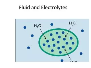
Fluid • Approximately 60% of typical adult is fluid • Varies with age, body size, gender • Intracellular fluid • Extracellular fluid • Intravascular: the fluid within the blood vessels e.g. plazma • Interstitial: fluid surrounds the cell (e.g. lymph • Transcellular: cerebrospinal, pericardial, synovial, intraocular, pleural, sweat and digestive secretion • “Third spacing”: loss of ECF into space that does not contribute to equilibrium
Electrolytes • Active chemicals that carry positive (cations), negative (anions) electrical charges • Major cations: sodium, potassium, calcium, magnesium, hydrogen ions • Major anions: chloride, bicarbonate, phosphate, sulfate, and proteinate ions • Electrolyte concentrations differ in fluid compartments
Regulation of Fluid • Movement of fluid through capillary walls depends on • Hydrostatic pressure: exerted on walls of blood vessels • Osmotic pressure: exerted by protein in plasma • Direction of fluid movement depends on differences of hydrostatic, osmotic pressure
Regulation of Fluid • Osmosis: area of low solute concentration to area of high solute concentration • Diffusion: solutes move from area of higher concentration to one of lower concentration • Filtration: movement of water, solutes occurs from area of high hydrostatic pressure to area of low hydrostatic pressure • Active transport: physiologic pump that moves fluid from area of lower concentration of one of higher concentration
Active Transport • Physiologic pump that moves fluid from area of lower concentration to one of higher concentration • Movement against concentration gradient • Sodium-potassium pump: maintains higher concentration of extracellular sodium, intracellular potassium • Requires adenosine (ATP) for energy
Question • Tell whether the following statement is true or false: • Osmosis is the movement of a substance from an area of higher concentration to one of lower concentration.
Answer • False. • Rationale: Diffusion is the movement of a substance from an area of higher concentration to one of lower concentration. The concentration of dissolved substances draws fluid in that direction. Osmosis is the movement of fluid, through a semipermeable membrane, from an area of low solute concentration to an area of high solute concentration until the solutions are of equal concentration.
Routes of Gains and Losses • Gain • Dietary intake of fluid, food or enteral feedingIngested fluid (60%) and solid food (30%) • Metabolic water or water of oxidation (10%) • Parenteral fluids
Routes of Gains and Losses (cont’d) • Loss • Kidney: urine output • Skin loss: sensible, insensible losses • Lungs • GI tract • Other • Urine (60%) and feces (4%) • Insensible losses (28%), sweat (8%)
Chapter 26: Fluid, Electrolyte, and Acid-Base Balance Water Intake and Output Figure 26.4
Question • What is the average daily urinary output in an adult? • 0.5 L • 1.0 L • 1.5 L • 2.5 L
Answer • C. 1.5 L • Rationale: Vital to the regulation of fluid and electrolyte balance, the kidneys normal filter 170 L of plasma every day in the adult, while excreting only 1.5 L of urine.
Regulation of Water Intake • The hypothalamic thirst center is stimulated: • By a decline in plasma volume of 10%–15% • By increases in plasma osmolality of 1–2% • Via baroreceptor input, angiotensin II, and other stimuli
Thirst is reduced as soon as we begin to drink water • Feedback signals that inhibit the thirst centers include: • Moistening of the mucosa of the mouth and throat • Activation of stomach and intestinal stretch receptors
Regulation of Water Output • Obligatory water losses include: • Insensible water losses from lungs and skin • Water that accompanies undigested food residues in feces • Obligatory water loss reflects the fact that: • Kidneys excrete 900-1200 mOsm of solutes to maintain blood homeostasis • Urine solutes must be flushed out of the body in water
Influence and Regulation of ADH • Water reabsorption in collecting ducts is proportional to ADH release • Low ADH levels produce dilute urine and reduced volume of body fluids • High ADH levels produce concentrated urine • Hypothalamic osmoreceptors trigger or inhibit ADH release • Factors that specifically trigger ADH release include prolonged fever; excessive sweating, vomiting, or diarrhea; severe blood loss; and traumatic burns
Mechanisms and Consequences of ADH Release Figure 26.6
Fluid Volume Imbalances • Fluid volume deficit (FVD): hypovolemia • Fluid volume excess (FVE): hypervolemia
Fluid Volume Deficit • Loss of extracellular fluid exceeds intake ratio of water • Electrolytes lost in same proportion as they exist in normal body fluids • Dehydration: loss of water along with increased serum sodium level • May occur in combination with other imbalances
Fluid Volume Deficit (cont’d) • Dehydration • Causes: fluid loss from vomiting, diarrhea, GI suctioning, sweating, decreased intake, inability to gain access to fluid • Risk factors: diabetes insipidus, adrenal insufficiency, osmotic diuresis, hemorrhage, coma,
Fluid Volume Deficit (cont’d) • Manifestations: rapid weight loss, decreased skin turgor, oliguria, concentrated urine, postural hypotension, rapid weak pulse, increased temperature, cool clammy skin due to vasoconstriction, lethargy, thirst, nausea, muscle weakness, cramps • Laboratory data: elevated BUN in relation to serum creatinine, increased hematocrit • Serum electrolyte changes may occur
Fluid Volume Deficit (cont’d) • Medical management: provide fluids to meet body needs • Oral fluids • IV solutions (isotonic electrolytes solution; e.g. ringer lactate solution or 0.9 sodium chloride)
Fluid Volume Deficit - Nursing Management • I&O, VS • Monitor for symptoms: skin and tongue turgor, mucosa, UO, mental status • Measures to minimize fluid loss • Oral care • Administration of oral fluids • Administration of parenteral fluids
Question • What is a major indicator of extracellular FVD? • Full and bounding pulse • Drop in postural blood pressure • Elevated temperature • Pitting edema of lower extremities
Answer • B. Drop in postural blood pressure • Rationale: FVD signs and symptoms include acute weight loss; decreased skin turgor; oliguria; concentrated urine; orthostatic hypotension due to volume depletion; a weak, rapid heart rate; flattened neck veins; increased temperature; thirst; decreased or delayed capillary refill; decreased central venous pressure; cool, clammy, pale skin related to peripheral vasoconstriction; anorexia; nausea; lassitude; muscle weakness; and cramps. Clinical manifestations of FVE result from expansion of the ECF and include edema, distended neck veins, and crackles (abnormal lung sounds).
Fluid Volume Excess • Due to fluid overload or diminished homeostatic mechanisms • Risk factors: heart failure, renal failure, cirrhosis of liver • Contributing factors: excessive dietary sodium or sodium-containing IV solutions • Manifestations: edema, distended neck veins, abnormal lung sounds (crackles), tachycardia, increased BP, pulse pressure and CVP, increased weight, increased UO, shortness of breath and wheezing • Medical management: directed at cause, restriction of fluids and sodium, administration of diuretics
Disorders of Water Balance: Edema • Atypical accumulation of fluid in the interstitial space, leading to tissue swelling • Caused by anything that increases flow of fluids out of the bloodstream or hinders their return • Factors that accelerate fluid loss include: • Increased blood pressure, capillary permeability • Incompetent venous valves, localized blood vessel blockage • Congestive heart failure, hypertension, high blood volume
Edema • Hindered fluid return usually reflects an imbalance in colloid osmotic pressures • Hypoproteinemia – low levels of plasma proteins • Forces fluids out of capillary beds at the arterial ends • Fluids fail to return at the venous ends • Results from protein malnutrition, liver disease, or glomerulonephritis
Chapter 26: Fluid, Electrolyte, and Acid-Base Balance Edema • Blocked (or surgically removed) lymph vessels: • Cause leaked proteins to accumulate in interstitial fluid • Exert increasing colloid osmotic pressure, which draws fluid from the blood • Interstitial fluid accumulation results in low blood pressure and severely impaired circulation
Fluid Volume Excess - Nursing Management • I&O and daily weights; assess lung sounds, edema, other symptoms; monitor responses to medications- diuretics • Promote adherence to fluid restrictions, patient teaching related to sodium and fluid restrictions • Monitor, avoid sources of excessive sodium, including medications • Promote rest • Semi-Fowler’s position for orthopnea • Skin care, positioning/turning
Electrolyte Imbalances • Sodium: hyponatremia, hypernatremia • Potassium: hypokalemia, hyperkalemia • Calcium: hypocalcemia, hypercalcemia • Magnesium: hypomagnesemia, hypermagnesemia • Phosphorus: hypophosphatemia, hyperphosphatemia • Chloride: hypochloremia, hyperchloremia
Hyponatremia • Serum sodium less than 135 mEq/L • Causes: adrenal insufficiency, water intoxication, SIADH (Syndrome of inappropriate ADH secretion) or losses by vomiting, diarrhea, sweating, diuretics • Manifestations: poor skin turgor, dry mucosa, headache, decreased salivation, decreased BP, nausea, abdominal cramping, neurologic changes • Medical management: water restriction, sodium replacement • Nursing management: assessment and prevention, dietary sodium and fluid intake, identify and monitor at-risk patients, effects of medications (diuretics, lithium)
Hypernatremia • Serum sodium greater than 145mEq/L • Causes: excess water loss, excess sodium administration, diabetes insipidus, heat stroke, hypertonic IV solutions • Manifestations: thirst; elevated temperature; dry, swollen tongue; sticky mucosa; neurologic symptoms; restlessness; weakness • Note: thirst may be impaired in elderly or the ill • Medical management: hypotonic electrolyte solution or D5W • Nursing management: assessment and prevention, assess for OTC (Over the counter drugs) sources of sodium, offer and encourage fluids to meet patient needs, provide sufficient water with tube feedings
Hypokalemia • Below-normal serum potassium (<3.5 mEq/L), may occur with normal potassium levels with alkalosis due to shift of serum potassium into cells • Causes: GI losses, medications, alterations of acid-base balance, hyperaldosterism, poor dietary intake • Manifestations: fatigue, anorexia, nausea, vomiting, dysrhythmias, muscle weakness and cramps, paresthesias, glucose intolerance, decreased muscle strength and DTRs • Medical management: increased dietary potassium, potassium replacement, IV for severe deficit • Nursing management: assessment, severe hypokalemia is life-threatening, monitor ECG and ABGs, dietary potassium, nursing care related to IV potassium administration
Hyperkalemia • Serum potassium greater than 5.0 mEq/L • Causes: usually treatment related, impaired renal function, hypoaldosteronism, tissue trauma, acidosis • Manifestations: cardiac changes and dysrhythmias, muscle weakness with potential respiratory impairment, paresthesias, anxiety, GI manifestations • Medical management: monitor ECG, limitation of dietary potassium, cation-exchange resin (Kayexalate), IV sodium bicarbonate , IV calcium gluconate, regular insulin and hypertonic dextrose IV, -2 agonists, dialysis
Hyperkalemia (cont’d) • Nursing management: assessment of serum potassium levels, mix IVs containing K+ well, monitor medication affects, dietary potassium restriction/dietary teaching for patients at risk • Hemolysis of blood specimen or drawing of blood above IV site may result in false laboratory result • Salt substitutes, medications may contain potassium • Potassium-sparing diuretics may cause elevation of potassium • Should not be used in patients with renal dysfunction
Question • Tell whether the following statement is true or false: • The ECG change that is specific to hyperkalemia is a peaked T wave.
Answer • True. • Rationale: The ECG changes that are specific to hyperkalemia are peaked T wave; wide, flat P wave; and wide QRS complex. The ECG changes that are specific to hypokalemia are flatted T wave and the appearance of a U wave.
Hypocalcemia • Serum level less than 8.5 mg/dL, must be considered in conjunction with serum albumin level • Causes: hypoparathyroidism, malabsorption, pancreatitis, alkalosis, massive transfusion of citrated blood, renal failure, medications, other • Manifestations: tetany, circumoral numbness, paresthesias, hyperactive DTRs, Trousseau’s sign, Chovstek's sign, seizures, respiratory symptoms of dyspnea and laryngospasm, abnormal clotting, anxiety
Hypocalcemia (cont’d) • Medical management: IV of calcium gluconate, calcium and vitamin D supplements; diet • Nursing management: assessment, severe hypocalcemia is life-threatening, weight-bearing exercises to decrease bone calcium loss, patient teaching related to diet and medications, and nursing care related to IV calcium administration