Download
1 / 116
1.16k likes | 1.23k Views
Explore the balance and distribution of water, osmolality, and concentrations of solutions in the body. Learn about the movement of water, osmosis, and other mechanisms affecting fluid regulation. Dive into the functions of the kidneys, hormonal control, and regulation of body fluids for optimal health.

E N D
Fluid and Electrolytes CSON Spring 2009 PREPARED BY CARLA HILTON, MSN, RN PRESENTED AND REVISED BY REBECCA POWERS, MSN, RN 15 questions from all of powers’ stuff…
Water Balance = Homeostasis • Water in the body is used to or for: • Transporting nutrients & oxygen to cells • Removing waste from cells • Provides medium in which electrolyte chemical reactions can occur • Regulation of body temperature • Lubricates joints and membranes • Provides medium for food digestion • liter of water weighs 2.2 lbs • The most accurate way to measure fluid status in a person is daily weights, not I&O!!!
Water Distribution • ICF: Intracellular fluid • ECF: Extracellular fluid (lymph system, interstitial fluid, intravascular fluid or plasma) • TCF: Transcellular fluid (cerebral spinal fluid, fluid in joints, GI tract, and peritoneal fluid) • Third spacing: (a condition where fluid accumulates in a pocket that isn’t really serving a purpose. Acieties (sp?)- where fluid hangs out in your abd. The fluid is coming from somewhere else.) • More fluid in intracellular than anywhere else in the body!
Osmolarity / Osmolality • Osmole: • the amount of substance that dissociates in solution to form one mole of osmotically active particles • Concentration of solution measured in osmoles
Osmolarity / Osmolality • Osmolality is measured in milliOsmols/Kg (mOsm/Kg) • Osmolarity is measured in milliOsmols/L (mOsm/L) • Evaluates serum and urine in clinical practice • Normal: serum osmolality 275 – 295 mOsm/K • Lality= total volume will equal 1 L plus the amount of volume taken up by the solids! The koolaid and water equal a L • Larity= volume is going to be less than 1 L. The koolaid minus the water.
Concentrations of Solutions • Isotonic: Same osmolarity as blood plasma…no osmotic “pull” • Hypotonic: Less concentration than blood plasma…lower osmotic pressure • Hypertonic: More concentration than blood plasma….higher osmotic pressure
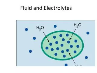
Movement of Water • Intracellular & extracellular approximately same osmolality • Solvent (water) and solutes (electrolytes) move across selectively permeable membranes (compartments) in the body (the bigger the particle, the slower they move, and they may need a little boost…)
Review of Terms • Osmosis • Diffusion • Active transport • Passive transport • Filtration • Hydrostatic pressure
Osmosis Review • Movement of water only • Speed of movement affected by: • temperature of fluid • concentration of fluid • electrical charge of particles in solution • The higher the solute concentration, the greater the osmotic pressure is.
Other Mechanisms of Movement • Diffusion: Solute (or gas) moves from area of higher concentration to area of lower concentration • Facilitated diffusion: Solute moves against concentration gradient (passive transport) • Active transport: Solute moved against concentration gradient using ENERGY
Active Transport • Na+/K+ pump: Maintains the higher concentrations of extracellular Na+ and intracellular K+ • In the cell, K is King. i.e. K is the major cation of the cell, Na is outside the cell.
Continued • Filtration: solutes & solvent move together in response to fluid pressure; moves from area of high pressure (hydrostatic pressure) to area of low pressure • Hydrostatic pressure: The force within a fluid compartment (as in the vascular system) The pressure that forces the fluid out of your capillaries. • Colloidal Osmotic Pressure – pulls it back into the capillaries.
Regulation of Body Fluids • Intake: osmoreceptors senseosmolality of serum, signals the hypothalamus, stimulates thirst • Impact on intake: Age (decreases desire to drink), conciousness, ability to take in fluids • Output: kidneys, lungs, GI tract, skin • Sensible: measurable….urine output, excessive perspiration, diarrhea, vomiting • Insensible: immeasurable…normal perspiration, normal breathing • Output for adults should be one mL/kg (of body weight) an hour
Role of the Kidneys • Filter approx 180 Liters of blood per day; GFR (glomerular filtration rate) • Produces urine between 1-2 Liters/day • If loss of 1% to 2% of body water, will conserve water by reabsorbing more water from filtrate; urine will be more concentrated • If gain of excess body water, will excrete more water from filtrate; urine will be more diluted
Hormonal Control • Antidiuretic hormone (ADH): Prevents diuresis; “water saving” • Question: Osmoreceptors sensing a/an increase in osmolality will cause the release of ADH • ADH acts on kidneys via the renal tubules. Makes them more permeable to water. The water will move from the tubes back into your body.
Hormonal Control • RAA (Renin-angiotensin-aldosterone):cascade initiated by decrease in renal perfusion or low Na+ • If extracellular volume is decreased renal perfusion decreases renin secreted by kidneys renin acts to produce angiotensin I which then converts to angiotensin II results in massive vasoconstriction increases renal arterial perfusion and causes increased thirst, a release of aldosterone (causes the retention of Na and Water)
Hormonal Control • Aldosterone: • Angiotensin II causes the adrenal gland to release aldosterone • Aldosterone causes the kidneys to retain Na+ and water • Volume regulator….released if Na+ is low and K+ is high; increases reabsorption of Na+ (where salt goes, water follows) and the excretion of K+
ANP • Atrial Natriuretic Peptide: (ANP): secreted from atrial cells of heart (in response to too much volume in the blood) • acts as diuretic • inhibits thirst mechanism • suppresses the RAA cascade
Thirst Mechanism • Regulated by the hypothalamus • Stimulates thirst: • increased osmolality of ECF • decreased ECF • dry mucous membranes • Causes: eating salty foods, inadequate intake, excessive water loss
Pressure Sensors Baroreceptors: Nerve receptors that sense pressure in blood vessels (think barometer measures pressure in the atmosphere, this measures pressure in the blood vessels) • Low pressure: sensors in the cardiac atria; stimulate SNS (sympathetic nervous system) & inhibits PSNS (parasympathetic nervous system) (sns will increase heart rate and BP) • High pressure: sensors in the aortic arch, carotid sinus, and the juxtaglomerular apparatus in the kidney; stimulates PSNS and inhibits the SNS (psns will decrease your heart rate and lower BP)
Pressure Sensors • Osmorecptors: Sense Na+ concentration • Positioned on surface of hypothalamus • Increase in Na+ concentration: stimulates release of ADH • Decrease in Na+ concentration: inhibits release of ADH
Electrolytes • Minerals and salts: electrolytes • Cations: Positively charged; sodium, potassium, calcium, magnesium • Major cation in ECF is sodium • Anions: Negatively charged; chloride, bicarbonate, sulfate • Major cation in ICF is potassium
HyponatremiaUsually loss of Na w/o loss of fluid • Causes • Salt wasting fr. Kidney • Adrenal insufficiency • GI losses • Profuse sweating • Diuretics • SIADH • Syndrome of inappropriate Anti-Diruetic Hormone • Inadequate Na intake • Physical Exam • Apprehension • Personality change • Postural hypotension • Tachycardia • Convulsions/coma • NV&D • Anorexia
Hyponatremia cont’d • Labs • Serum Na+below 135 mEq/L • Serum Osmolality below 280 mOsm/kg • Urine specific gravity below 1.010 • Treatment • Restrict water • Sodium replacement
Hypernatremia • Causes • ingestion of salt • Iatrogenic (we caused it) • aldosterone • Water deprivation • Signs & Sxms • Thirst, sticky tongue • Dry, flushed skin • Fever • Convulsions, irritability
Hypernatremia cont’d • Labs • Serum Na+above 145 mEq/L • Serum Osmolality above 295 mOsm/kg • Urine specific gravity above 1.030 • Treatment • Hypotonic IV solution • or D5W
Urine Na+ Studies • Urine Na+ • Assesses volume status • Aids in diagnosing hyponatremia & acute renal failure • Random normal range = 50 -130 mEq/L • 24 hour = 75-200 mEq/L
Hypokalemia • Causes • Diuretics that “waste” potassium • D, V, & gastric suction • aldosterone • Polyuria, sweating • Iatrogenic – K+ poor solutions • Signs & Sxms • Weakness, fatigue • muscle tone • Hypoactive bowel sounds and distention • Weak, irregular pulse • Paresthesias • SOMETHING ABOUT CARDIAC FUNCTION
Hypokalemia cont’d • Labs • K+below 3.5 mEq/L • ECG abnormalities • Treatment • Oral K+ or IV solution w/K+ • Increased dietary K+
Hyperkalemia • Causes • Renal failure • Fluid vol. deficit • Massive cellular injury (trauma/burns) • Iatrogenic • Potassium “sparing” diuretics • Addison’s disease • Signs & Sxms • Anxiety • Dysrrhythmias • Paresthesia (numbness, pins & needles feeling) • Weakness • Diarrhea
Hyperkalemia cont’d • Labs • Serum K+above 5.0 mEq/L. • ECG abnormalities – can lead to arrest (if too high or too low) • Treatment • Kayexalate • IV Na+ bicarb • IV Ca+ gluconate • Regular insulin and hypertonic dextrose IV • Limit via diet • Possible dialysis
Hypocalcemia • Causes • Rapid admin of blood w citrate • Hypoalbuminemia • Hypoparathyroidism • Vit. D deficiency • Pancreatitis • Stuff that relates back to preexisting conditions • Signs & Sxms • Numbness, tingling of fingers & mouth • Hyperactive reflexes • Tetany- a muscle contraction that stays contracted • Muscle cramps • Pathological fractures
Hypocalcemia cont’d • Labs • Serum Ca++below 4.5 mEq/L • ECG abnormalities • Treatment • Increase dietary intake • IV calcium gluconate • Ca+ & vit D supplements
Hypercalcemia • Causes • Hyperparathyroidism • Osteometastasis • Paget’s disease • Osteoporosis • Prolonged immobilization • Signs & Sxms • Anorexia, N & V • Weakness, lethargy • Low back pain (stones) • Decreased LOC • Personality changes • Cardiac arrest
Hypercalcemia cont’d • Labs • Serum Ca++above 5.5 mEq/L • X-rays showing osteoporosis • Stones & BUN / creatinine fr. FVD or renal damage • Treatment • Lasix (diuretic) • Increased fluids
Hypomagnesemia • Causes • Inadequate intake • Alcohol, Malnutrition • Inadequate absorption • V&D, Gastric aspirate • Fistulas, Sm. Bowel • Loss fr. Diuretics • Polyuria • Signs & Sxms • Tremors • Hyperactive deep tendon reflexes • Confusion • Dysrhythmias
Hypomagnesemia cont’d • Labs • Serum Mg++below 1.5 mEq/L • Treatment • Mag sulfate IV • Oral replacement • Increase dietary intake
Hypermagnesemia • Causes • Renal failure • Excess intake of magnesium • Signs & Sxms • Most frequently seen in acute • Hypoactive deep tendon reflexes & drowsiness • Decreased depth and rate of resp. • Hypotension • flushing
Hypermagnesemia cont’d • Labs • Serum Mg++ levels above 2.5 mEq/L • Treatment • IV calcium gluconate • Loop diuretics • NS or LR IV solutions • Dialysis
Additional Lab Data • Hematocrit • Measures the volume % of RBC’s in whole blood • Normal: M = 40-50%; F = 37-47% • Increases with dehydration (hemoconcentration) • Decreases with overhydration (hemodilution)
Hematocrit & Fluid Volume StatusFrom “Fluids & Electrolytes Made Incredibly Easy” 4th ed. Fluids & Electrolytes Made Incredibly Easy
Lab Data (cont’d) • Blood urea nitrogen (BUN) • Measures kidney function • Normal range: 7-20mg/dL • Varies with protein intake, fever, dehydration, GI bleeding, liver failure, etc.
Lab Data (cont’d) • Creatinine • End product of muscle metabolism • Better indicator of renal function than BUN • Doesn’t vary w protein intake or metabolic state • Normal range: 0.7-1.5mg/dL in 24 hr urine collection • Serum: adult female: 0.5 to 1.1mg/dL adult male: 0.6 to 1.2mg/dL
Lab Data (cont’d) • Urine Specific Gravity • Measures ability of kidney to excrete or conserve water • Normal range = 1.010 - 1.025 • Increased S.G.= concentrated urine • Decreased S.G.= dilute urine
Lab Data (cont’d) • Serum Osmolarity • Most accurate for kidney function • Remember norm? • 280-295 mOsm/L • Measured directly through blood • Indirectly using Serum Osmolarity Formula
Fluid Imbalances • Isotonic • Deficit – water, electrolytes and solutes lost in equal proportions to body solutions • Excess – water, electrolytes and solutes gained in equal proportions to body solution • FVD - fluid volume deficit-HYPOVOLEMIA • FVE - fluid volume excess-HYPERVOLEMIA