Download
1 / 26
280 likes | 458 Views
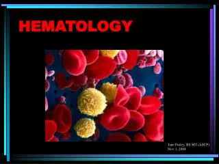
Discover the composition of blood elements, different stages of hematopoiesis, and disorders related to red and white blood cells. Learn about hemostasis and platelet function.

E N D
Hematology Introduction Organization of blood and blood forming organs CLS245
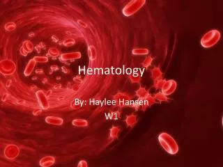
What is hematology? • Hematology is the study of blood which is composed of plasma (~55%), and the formed elements which are: • The erythrocytes (RBCs) (~45%) • Contain hemoglobin • Function in the transport of O2 and CO2 • The Leukocytes (WBCs) and platlets (thrombocytes) (~1%) • Leukocytes are involved in the body’s defense against the invasion of foreign antigens. • Platelets are involved in hemostasis which forms a barrier to limit blood loss at an injured site.
What is hematology? • Hematology is primarily a study of the formed cellular elements. • Alterations in the formed elements in the blood are usually a result of disease rather than being the primary cause of disease.
WHAT IS HEMATOLOGY? • Hematopoiesis is a term describing the formation and development of blood cells.
Hematopoiesis • Mesoblastic stage • Nucleated RBCs - Yolk sac and Mesothelial layers of the placenta – 3rd week • Hepatic stage • Primitive erythroblast > migrate to liver and spleen (6wks) – includes other myeloid and lymphoid cells. • Myeloid stage • From the third month onwards - the bone marrow gradually becomes the principal source of the RBCs • Last month (9th month) – Bone marrow exclusively
Hematopoiesis • At birth the bone cavities are the only site of hematopoietic activity - Starts at the 7th months • Yolk sac > fetal liver/spleen > BM
Relative rates of red blood cell production in the bone marrow of different bones at different ages.
Hematopoiesis Hematopoietic stem cell pool • Pluripotential - multipotential stem cells (MSCs) • morphologically identical • Continuous self-renewal • Unipotential -committed progenitor cells • Restricted to single cell line • No self-renewal • Lymphoid or myeloid
mature red cell (RBC) • Size: 6 to 8 μm • Appearance: Disk-shaped cell filled with hemoglobin, having an area of central pallor of 1 to 3 μm
Mature RBCs • Size: 6 to 8 μm, having an area of central pallor of 1 to 3 μm, • Biconcave discs, no nucleus, essentially no organelles. • Filled with hemoglobin (Hb), a protein that functions in gas transport. • Contain the plasma membrane protein spectrin and other proteins that give erythrocytes their flexibility and allow them to change shape as necessary.
Hemoglobin • HB is a highly specialized intracellular erythrocyte protein, responsible for transport O2 from the lung to the tissues and CO2 from tissues to the lung. • It is a tetramer, consisting of two pairs of similar polypeptide chains called globins, To each of the four chains is attached a prosthetic group, heme, a complex of iron and protoporphyrin.
Red blood cells disorders Anemia Is a reduction of circulating red blood cells, hemoglobin, and packed cell volume to a level lower than the normal range.
It can be classified according to the etiology, or to the morphological appearance of the RBCs under light microscope. 1- Morphological classification: • Microcytic hypochromic anemias: Ex. Iron deficiency anemia • Macrocytic normochromic anemias: Ex. Megablastic anemia • Normocytic normochromic anemias: Ex. Sickle cell anemia 2- Etiological classification: • This classification is based on the cause of anemia. • Increase destruction. • Decrease production. • Blood loss (hemorrhage).
White blood cells disorders • Leukemia = lymphoid and myeloid malignant bone marrow neoplasms • when abnormal cells are seen in both the bone marrow and the peripheral blood • Lymphoma = abnormal proliferation of lymphoid cells within the lymphatic tissue or lymph nodes Leukemia: • Acute leukemia = rapid onset with abnormal expansion of immature cells • Chronic leukemia = slow progression with abnormal expansion of mature cells • Divided by two cell types: myeloid and lymphoid • Acute myeloid leukemia (AML), or chronic myeloid leukemia (CML) • Acute lymphoid leukemia (ALL), or chronic lymphoid leukemia (CLL)
HEMOSTASIS • Hemostasis can be defined as: the stoppage of bleeding from the blood vessels • It is a complex process in which several factors work together or in sequence to stop the flow of blood from an injured blood vessel. • The five major components of normal hemostasis are blood vessels constriction, platelets, coagulation factors, coagulation inhibitors and fibrinolysis.
PLATELETS • Platelets are small fragments of cytoplasm derived from megakaryocytes. On average they are 1.5–3.5 μm in diameter but may be larger in some diseases.
Hemostasis Disorders EX. Immune thrombocytopenic purpura (ITP) is one of the most common autoimmune disorders. ITP is caused by autoantibodies to platelets. The antigenic target in most patients appears to be the platelet GP IIb/IIIa complex. Platelets with antibodies on their surface are trapped in the spleen, where they are efficiently removed by splenic macrophages.
Hematology Lab Tests 1- The complete blood count, or CBC lists a number of many important values. Typically, it includes the following: • White blood cell count (WBC or leukocyte count) • WBC differential count • Red blood cell count (RBC or erythrocyte count) • Hematocrit (Hct) • Hemoglobin (Hbg) • Mean corpuscular volume (MCV) • Mean corpuscular hemoglobin (MCH) • Mean corpuscular hemoglobin concentration (MCHC) • Red cell distribution width (RDW) • Platelet count • Mean Platelet Volume (MPV)
Hematology Lab Tests 2- Sickle cell test 3- peripheral blood film 4- Erythrocyte Sedimentation Rate nonspecific test “ look for general infection only “ 5- Hb Electrophoresis 6- Coagulation tests “ PT, APTT, TT, D-Dimer” 7- Bone marrow aspiration.