Understanding Miniature End-Plate Potentials at the Neuromuscular Junction
This article explores the anatomy of the neuromuscular junction (NMJ) with a focus on miniature end-plate potentials (MEPPs). Scientists recorded end-plate potentials (EPPs) induced by nerve stimulation and noted spontaneous depolarizations resembling miniature EPPs. These MEPPs can be evoked under specific conditions of low Ca²⁺ and high Mg²⁺. The quantum hypothesis suggests that MEPPs are the fundamental units of synaptic transmission, with each quantal event representing a fixed amount of neurotransmitter, about 7000 acetylcholine molecules at the NMJ.

Understanding Miniature End-Plate Potentials at the Neuromuscular Junction
E N D
Presentation Transcript
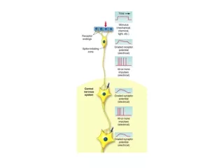
Synaptic Communication 5 March 2012
The story of MEPPs Scientists recorded membrane potentials from the end-plate (EPPs), evoked by nerve stimulation They also observed spontaneous depolarizations of approximately 1mV These looked like miniature EPPs that they could evoke by nerve stimulation They could evoke these MEPPs, or “minis,” under conditions that limited ACh release (low Ca2+, high Mg2+)
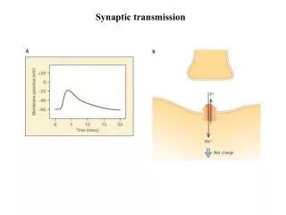
MEPPs: Miniature End Plate Potentials A: Spontaneous MEPPs recorded from frog muscle in normal Ringer's solution. Arrows indicate 1 mV, 20 ms. B: Response recorded from end-plate region of frog muscle to repeated nerve stimulation at arrows. Low-Ca Ringer's solution with neostigmine bromide (1 g/ml). Note stepwise fluctuation in EPP's on successive trials. Voltage scale, 1 mV. Images From Fatt & Katz (1956)
EPPs vs. MEPP Amplitudes From Martin and Brodie, 1956
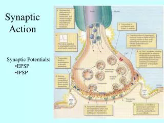
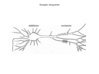
Quantum Hypothesis • Hypothesis: Single quantal events (minis) are the building blocks for the synaptic potentials evoked by stimulation • Each quanta is a packet of neurotransmitter, and each quanta contains the same amount of NT • How much? • Varies; about 7000 ACh molecules at the NMJ