Download
1 / 121
1.21k likes | 1.23k Views
Nervous System. The master controlling and communicating system of the body Functions Sensory input – monitoring stimuli Integration – interpretation of sensory input Motor output – response to stimuli. Nervous System. Figure 11.1. Organization of the Nervous System.

E N D
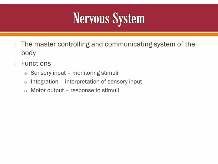
Nervous System • The master controlling and communicating system of the body • Functions • Sensory input – monitoring stimuli • Integration – interpretation of sensory input • Motor output – response to stimuli
Nervous System Figure 11.1
Organization of the Nervous System • Central nervous system (CNS) • Brain and spinal cord • Integration and command center • Peripheral nervous system (PNS) • Paired spinal and cranial nerves • Carries messages to and from the spinal cord and brain
Peripheral Nervous System (PNS): Two Functional Divisions • Sensory (afferent) division • Sensory afferent fibers – carry impulses from skin, skeletal muscles, and joints to the brain • Visceral afferent fibers – transmit impulses from visceral organs to the brain • Motor (efferent) division • Transmits impulses from the CNS to effector organs
Motor Division: Two Main Parts • Somatic nervous system • Conscious control of skeletal muscles • Autonomic nervous system (ANS) • Regulates smooth muscle, cardiac muscle, and glands • Divisions – sympathetic and parasympathetic
Histology of Nerve Tissue • The two principal cell types of the nervous system are: • Neurons – excitable cells that transmit electrical signals • Supporting cells – cells that surround and wrap neurons
Supporting Cells: Neuroglia • The supporting cells (neuroglia or glial cells): • Provide a supportive scaffolding for neurons • Segregate and insulate neurons • Guide young neurons to the proper connections • Promote health and growth
Astrocytes • Most abundant, versatile, and highly branched glial cells • They cling to neurons and their synaptic endings, and cover capillaries
Astrocytes • Functionally, they: • Support and brace neurons • Anchor neurons to their nutrient supplies • Guide migration of young neurons • Control the chemical environment
Microglia and Ependymal Cells • Microglia – small, oval cells with spiny processes • Phagocytes that monitor the health of neurons • Ependymal cells – range in shape from squamous to columnar • They line the central cavities of the brain and spinal column Microglia Ependymal Cells
Oligodendrocytes, Schwann Cells, and Satellite Cells • Oligodendrocytes – branched cells that wrap CNS nerve fibers with myelin • Schwann cells (neurolemmocytes) – surround fibers of the PNS with myelin • Satellite cells surround neuron cell bodies with ganglia Oligodendrocytes Schwann Cells Satellite Cells
Neurons (Nerve Cells) • Structural units of the nervous system • Composed of a body, axon, and dendrites • Long-lived, amitotic, and have a high metabolic rate • Their plasma membrane function in: • Electrical signaling • Cell-to-cell signaling during development
Nerve Cell Body (Perikaryon or Soma) • Contains the nucleus and a nucleolus • Is the major biosynthetic center • Is the focal point for the outgrowth of neuronal processes • Has no centrioles (hence its amitotic nature) • Has well-developed Nissl bodies (rough ER) • Contains an axon hillock – cone-shaped area from which axons arise
Processes • Armlike extensions from the soma • Called tracts in the CNS and nerves in the PNS • There are two types: axons and dendrites
Dendrites of Motor Neurons • Short, tapering, and diffusely branched processes • They are the receptive, or input, regions of the neuron • Electrical signals are conveyed as graded potentials (not action potentials)
Axons: Structure • Slender processes of uniform diameter arising from the hillock • Long axons are called nerve fibers • Usually there is only one unbranched axon per neuron • Rare branches, if present, are called axon collaterals • Axonal terminal – branched terminus of an axon
Axons: Function • Generate and transmit action potentials • Secrete neurotransmitters from the axonal terminals • Movement along axons occurs in two ways • Anterograde — toward axonal terminal • Retrograde — away from axonal terminal
Myelin Sheath • Whitish, fatty (protein-lipoid), segmented sheath around most long axons • It functions to: • Protect the axon • Electrically insulate fibers from one another • Increase the speed of nerve impulse transmission
Myelin Sheath and Neurilemma: Formation • Formed by Schwann cells in the PNS • A Schwann cell: • Envelopes an axon in a trough • Encloses the axon with its plasma membrane • Has concentric layers of membrane that make up the myelin sheath • Neurilemma – remaining visible nucleus and cytoplasm of a Schwann cell
Myelin Sheath and Neurilemma: Formation Figure 11.5a–c
Nodes of Ranvier (Neurofibral Nodes) • Gaps in the myelin sheath between adjacent Schwann cells • They are the sites where axon collaterals can emerge
Unmyelinated Axons • A Schwann cell surrounds nerve fibers but coiling does not take place • Schwann cells partially enclose 15 or more axons
Axons of the CNS • Both myelinated and unmyelinated fibers are present • Myelin sheaths are formed by oligodendrocytes • Nodes of Ranvier are widely spaced • There is no neurilemma
Regions of the Brain and Spinal Cord • White matter – dense collections of myelinated fibers • Gray matter – mostly soma and unmyelinated fibers
Neuron Classification • Structural: • Multipolar — three or more processes • Bipolar — two processes (axon and dendrite) • Unipolar — single, short process
Neuron Classification • Functional: • Sensory (afferent) — transmit impulses toward the CNS • Motor (efferent) — carry impulses away from the CNS • Interneurons (association neurons) — shuttle signals through CNS pathways
Comparison of Structural Classes of Neurons Table 11.1.1
Comparison of Structural Classes of Neurons Table 11.1.2
Comparison of Structural Classes of Neurons Table 11.1.3
Neurophysiology • Neurons are highly irritable • Action potentials, or nerve impulses, are: • Electrical impulses carried along the length of axons • Always the same regardless of stimulus • The underlying functional feature of the nervous system
Electricity Definitions • Voltage (V) – measure of potential energy generated by separated charge • Potential difference – voltage measured between two points • Current (I) – the flow of electrical charge between two points • Resistance (R) – hindrance to charge flow • Insulator – substance with high electrical resistance • Conductor – substance with low electrical resistance
Electrical Current and the Body • Reflects the flow of ions rather than electrons • There is a potential on either side of membranes when: • The number of ions is different across the membrane • The membrane provides a resistance to ion flow
Role of Ion Channels • Types of plasma membrane ion channels: • Passive, or leakage, channels – always open • Chemically gated channels – open with binding of a specific neurotransmitter • Voltage-gated channels – open and close in response to membrane potential • Mechanically gated channels – open and close in response to physical deformation of receptors
Operation of a Gated Channel • Example: Na+-K+ gated channel • Closed when a neurotransmitter is not bound to the extracellular receptor • Na+ cannot enter the cell and K+ cannot exit the cell • Open when a neurotransmitter is attached to the receptor • Na+ enters the cell and K+ exits the cell
Operation of a Voltage-Gated Channel • Example: Na+ channel • Closed when the intracellular environment is negative • Na+ cannot enter the cell • Open when the intracellular environment is positive • Na+ can enter the cell
Gated Channels • When gated channels are open: • Ions move quickly across the membrane • Movement is along their electrochemical gradients • An electrical current is created • Voltage changes across the membrane
Electrochemical Gradient • Ions flow along their chemical gradient when they move from an area of high concentration to an area of low concentration • Ions flow along their electrical gradient when they move toward an area of opposite charge • Electrochemical gradient – the electrical and chemical gradients taken together
Resting Membrane Potential (Vr) • The potential difference (–70 mV) across the membrane of a resting neuron • It is generated by different concentrations of Na+, K+, Cl, and protein anions (A) • Ionic differences are the consequence of: • Differential permeability of the neurilemma to Na+ and K+ • Operation of the sodium-potassium pump
Resting Membrane Potential (Vr) Figure 11.8
Membrane Potentials: Signals • Used to integrate, send, and receive information • Membrane potential changes are produced by: • Changes in membrane permeability to ions • Alterations of ion concentrations across the membrane • Types of signals – graded potentials and action potentials
Changes in Membrane Potential • Changes are caused by three events • Depolarization – the inside of the membrane becomes less negative • Repolarization – the membrane returns to its resting membrane potential • Hyperpolarization – the inside of the membrane becomes more negative than the resting potential
Graded Potentials • Short-lived, local changes in membrane potential • Decrease in intensity with distance • Magnitude varies directly with the strength of the stimulus • Sufficiently strong graded potentials can initiate action potentials
Graded Potentials Figure 11.10
Graded Potentials • Voltage changes are decremental • Current is quickly dissipated due to the leaky plasma membrane • Only travel over short distances
Action Potentials (APs) • A brief reversal of membrane potential with a total amplitude of 100 mV • Action potentials are only generated by muscle cells and neurons • They do not decrease in strength over distance • They are the principal means of neural communication • An action potential in the axon of a neuron is a nerve impulse


