Download
1 / 40
400 likes | 539 Views
Chapter 11. Functional Organization of Nervous Tissue. Functions of the Nervous System. Collect Sensory Information: Sensory RECEPTORS detect stimuli Monitor internal and external stimuli/change Integration:

E N D
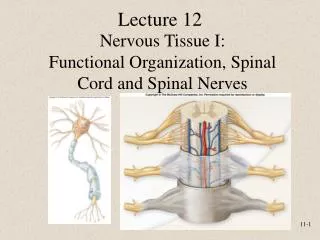
Chapter 11 Functional Organization of Nervous Tissue
Functions of the Nervous System • Collect Sensory Information: • Sensory RECEPTORS detect stimuli • Monitor internal and external stimuli/change • Integration: • Brain and/or spinal cord process sensory input/info and initiate (‘determine’) responses • Motor Output/Response • Stimulation of muscle and/or glands--EFFECTORS
Nervous system = PNS + CNS --Receives sensory input with receptors, sends/conducts sensory info to CNS, Carry commands to effectors from the CNS -- links the CNS to sensory information and effector Processes/integrates sensory information and generates responses
Organization of the Nervous System:General Pathway and Terms Receptor Sensory NS CNS Motor NS Effector
By receiving and integrating sensory input and signaling effectors the N.S. accomplishes the following: • Homeostasis. • Regulate and coordinate physiology • Response to internal conditions/change • Response to changes in external environment • Mental activity. • E.g., consciousness, thinking, memory, emotion
Divisions of the Nervous System • Sensory: • Any aspect of the N.S. involved with transmitting sensory Information • Somatic Sensory(afferent): • Sensory information from skin, muscle, or joints • Often conscious • Visceral Sensory (afferent): • Sensory information from internal organs (e.g., blood vessels, digestive organs, heart, etc….). • Typically not consciously aware of this sensory information
Divisions of the NS, continued • Motor: • Any part of the N.S. that transmits motor information • Somatic motor system: • fto skeletal muscles. • Voluntary. • Single neuron system from spinal cord to effector • Autonomic (visceral) motor system (ANS): • to smooth muscle, cardiac muscle, and certain glands. • Subconscious or involuntary control. • Two neurons from spinal cord to effector • Divisions of ANS • Sympathetic (SD)--Prepares body for physical activity/energy expenditure • Parasympathetic (PD)--Regulates resting or vegetative functions/ energy assimilation • Enteric--controls the digestive tract independently of the CNS, but is regulated/influenced by the SD & PD
More Complete Description of NS organization Not from your text, chapters don’t match up
NS Organization and Function: worked example 1. Stimulus: light receptors in eye send afferent impulses through sensory neurons (PNS). 2a. CNS integrates (processes) sensory information: you see a bear and are afraid, you decided to run away 2b. CNS generates instructions to run away. 3a. Efferent impulses travel through somatic motor neurons (PNS) to cause you skeletal muscle—effectors--to cause you to run away. 3b. Efferent impulses travel through visceral motor neurons (ANS/PNS) to smooth muscle (effector) and glands (effector) to do things like increase heart, respiratory rate, and dilate blood vessels so that your skeletal muscles have oxgyen and blood flow they need to function.
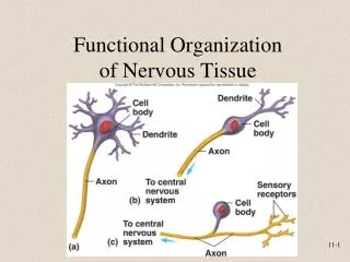
Cells of Nervous System • Neurons(nerve cells) receive stimuli and transmit action potentials • Major Parts • Cell body • Dendrites: • Axons: Transitive— generate output • Neuroglia or glial cells • Support and protect neurons Receptive--input
Parts of the Neuron • Cell Body (w/ Nucleus): • RECEPTIVE: receive input/stimuli from other cells or are stimulated directly • Graded potentials • Dendrites: • Many • RECEPTIVE: receives input/stimuli from other cells or are stimulated directly • Graded potentials • Axons. • 1 from cell body • Branches = collaterals. • synaptic knobs • With synaptic vesicles • Generates OUTPUT: Transmits impulses to other cells • Action potentials
Types of Neurons, Functional Categories:Types of neurons based on what they do • Sensoryorafferent: • Carry sensory information • Motororefferent: • Carry motor information • Interneuronsorassociationneurons: • Entirely contained within CNS from one neuron to another • Integration/processing
Figure 14.4 Types of Neurons, Structural Categories
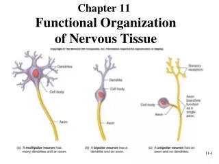
Types of Neurons, Structural Categories:Types of neurons based on shape and arrangement of parts • Multipolar: most neurons in CNS; motor neurons • Unipolar: single process that divides into two branches. Part that extends to the periphery has dendrite-like sensory receptors • e.g., sensory neurons of PNS • Bipolar: sensory in retina of the eye and nose
General Characteristics of Neurons • Do not divide* • Long, large • Requires effective transport Axoplasmic transport; elaborate transport mechanism to transport substances throughout the large cell • Requires/consumes large amount of energy • Ion pumps & gated channel proteins on PM • Enables electrical impulses (e.g., action potentials) • Moves ions • Requires/consumes large amounts of energy • Neurotransmitter release and reabsorption • E.g., sodium/potassium pump • High metabolic need (due to membrane and internal transport) • Many mitochondria • High oxygen demand so neurons and NS are very sensitive to lack of O2 • NS function/mental status are often earliest signs of dysfunction/loss of homeostasis *except in specific, limited areas of the brain
Electrical Signals • Electrical Activity of neurons is created by moving ions (mostly K+ and Na+) across the PM. • Involves changes in membrane permeability due to gated ion channels on the PM • Type types of electrical activity • graded (local) potentials: on dendrites and cell bodies • action potentials: on axons These Electrical Impulses: • Transfer of information/commands from one part of body to another
Synapses • Junction between two cells where electrical activity of one cell signals/influences electrical activity in another cell. • receptor—neuron • neuron—neuron • axon-dendrite/cell body • (axon-axon)—we will not discuss but they exist • neuron—effector (e.g., neuromuscular junction) • Synapses involve two cells • Presynaptic cells (proximal/before to the synaptic cleft) • Postsynaptic cells (distal/after the synaptic cleft) • Two types of synapses • Chemical Synapse (most common) • Electrical Synapse (less common)
Structure of a Chemical Synapse • Synaptic Knob (of presynaptic neuron) • calcium channels • Synaptic vessicles • Neurotransmitter • e.g., Ach, norepinephrine, dopamine, serotonin, glutamate • Synaptic cleft • Post synaptic cell: Post Synaptic membrane • Receptors (e.g., sodium gates)
Chemical Synapse Function • Electrical Impulses (action potentials) of presynaptic cell reaches synaptic knob • Calcium diffuses into knob • Causes release of neurotransmitter (by exocytosis) into cleft • Neurotransmitters diffuse across synaptic cleft • Neurotransmitters bind receptors of post synaptic cell causing: • Excitatory effects (EPSP) —promotes formation of electrical impulse (action potential) on postsynaptic cell • Inhibitory effects(IPSP)—inhibits formation of an electrical impulse (action potential) on postsynaptic cell
A B
Electrical Synapses • Gap junctions between pre- and postsynaptic cell allow electrical impulse (ion/charge) flow directing from one cell to another. • charges move throughprotein tubes—gap junctions--in cell membrane. • Found in cardiac muscle and many types of smooth muscle.
Neuroglia (glial cells) In PNS • Schwann cells • Satelite cells • Oligodendrocytes • Astrocytes • Microglia • Ependymal cells In CNS
Schwann cells(neurolemmocytes): Myelinated axon unmyelinated axon • Surround a portion of a single axon • (it takes many schwann cells to cover a single axon) • This “electrically insulates” the axon • Facilitates axon repair • Two arrangements: • Myelinated axons • Unmyelinated axons
Myelinated axons Myelin sheath = repeated wrapping, creating a layer of lipid & proteins rich PM over the axon surface. The Myelin sheath: “insulates” axons from one another, speeds transmission of action potential Not continuous Nodes of Ranvier Completion of Development of myelin sheaths at 1 yr. Degeneration of myelin sheaths occurs in multiple sclerosis and other disorders (de-myelination disorders). Myelinated Axons
Myelination PNS v. CNs Schwann Cells in PNS Oligodendrocytes in CNS
Myelination v non-myelinated myelinated unmyelinated
Rest/are located in in invaginations of Schwann cells Not wrapped around the axon Electrically insulates Don’t speed impulse Unmyelinated Axons
White vs. Gray Matter White matter: • Structures/regions rich with myelinated axons. • Carries electrical impulses from one place to another • e.g., columns (of spinal cord), tracts, nerves, Gray matter: • Areas of predominantly cell bodies, dendrites, neuroglia and/or unmyelinated axons, • Typically Integrative in function • E.g. spinal cord horns, cerebral cortex, dorsal root ganglia:.
Speed of Axon Conduction Two influences on speed of action potential • Myelination: • Myelinated axons are faster than unmyelinated • Diameter/thickness • The larger in diameter the axon the faster it carries an impulse
Structure of Nerves(including roots and rami) • Consist of • Axon bundles/nerve fibers • Schwann cells • Connective tissue • Blood vessels • Endoneurium: surrounds individual neurons • loose CT with capillaries (for neurons) • Perineurium: Surrounds fascicles • blood vessels Epineurium: surrounds the entire nerve • Dense CT
Satellite Cells of unipolar neuron • Satellite cells: surround neuron cell bodies in ganglia • Protects/regulates chemical environment around neuron • regulates nutrients
Astrocytes • covering surface of neurons & blood vessels • Regulate what substances reach the CNS from the blood (blood-brain barrier). • promote tight junctions to form blood-brain barrier • Regulate extracellular brain fluid composition • neurotransmitter movement/recycling • Assist in neuron repair • Guide development of neural connections • Hypothetically involved with processing (through Ca+ cycling)
Blood Brain Barrier (BBB) • Blood-brain barrier: The condition/situation in which the blood vessels (i.e., capillaries) of the CNS are relatively impermeable and prevent the passage of many substances that normally exit blood vessels. • Nervous system/neuron function in highly influenced by the chemical composition of the extracellular environment. • The BBB limits movement of substances between blood and brain and relies on astrocytes to regulate the extracellular environment of the CNS* • Barrier to drugs, e.g., chemotherapy drugs, others….
Neuroglia of CNS: Ependymal Cells • Line the ventricles and central canal. • Form/secrete cerebrospinal fluid (CSF) • Form part of the choroid plexuses (which make CSF)
Microglia • Phagocytic. • Defends against pathogens • Removes debri and dead tissue • Removes waste.