Download

1 / 94
970 likes | 1.06k Views
Explore the organization and functions of the nervous system, including divisions, types of neurons, and cellular components. Learn about neural transmission, potential changes, and impulse processing. Dive into the complexities of synapses and neural pathways.

E N D
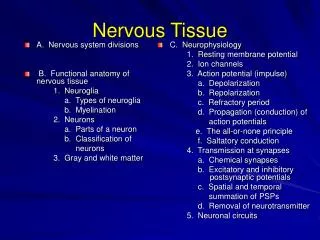
Divisions of the Nervous System • Central Nervous System • brain • spinal cord • Peripheral Nervous System • peripheral nerves • cranial nerves • spinal nerves 10-1
CNS PNS
Divisions of Peripheral Nervous System • Sensory Division • picks up sensory information and delivers it to the CNS • Motor Division • carries information to muscles and glands • Divisions of the Motor Division • Somatic – carries information to skeletal muscle • Autonomic – carries information to smooth muscle, cardiac muscle, and glands 10-4
Functions of Nervous System • Sensory Function • sensory receptors gather information • information is carried to the CNS • Motor Function • decisions are acted upon • impulses are carried to effectors • Integrative Function • sensory information used to create • sensations • memory • thoughts • decisions 10-6
Function of the Nervous System sensory input motor input sensory receptor effector integration
Types of Neuroglial Cells • Schwann Cells • peripheral nervous system • myelinating cell • Astrocytes • CNS • scar tissue • mop up excess ions, etc • induce synapse formation • connect neurons to blood vessels • Oligodendrocytes • CNS • myelinating cell • Ependyma • CNS • ciliated • line central canal of spinal cord • line ventricles of brain • Microglia • CNS • phagocytic cell 10-11
Neuron Structure 10-7
Typical Neuron dendrite cell body Myelin sheath Synapse axon
Neuron Membrane Outside cell Na+ -70mV K+ Inside cell
Myelination of Axons • White Matter • contains myelinated axons • Gray Matter • contains unmyelinated structures • cell bodies, dendrites 10-8
Nodes of Ranvier Schwann Cells Axon Myelin Sheath
Classification of Neurons • Bipolar • two processes • eyes, ears, nose • Unipolar • one process • ganglia • Multipolar • many processes • most neurons of CNS
Classification of Neurons • Sensory Neurons • afferent • carry impulse to CNS • most are unipolar • some are bipolar • Interneurons • link neurons • multipolar • in CNS • Motor Neurons • multipolar • carry impulses away from CNS • carry impulses to effectors 10-10
Resting Membrane Potential • inside is negative relative to the outside • polarized membrane • due to distribution of ions • Na+/K+ pump 10-14
Potential Changes • at rest membrane is polarized • threshold stimulus reached • sodium channels open and membrane depolarizes • potassium leaves cytoplasm and membrane repolarizes 10-15
Local Potential Changes • occur on membranes of dendrites and cell bodies • caused by various stimuli • chemicals • temperature changes • mechanical forces • if membrane potential becomes more negative, it has hyperpolarized • if membrane potential becomes more positive, it has depolarized • graded • summation can lead to threshold stimulus that starts an action potential 10-16
Action Potentials • nerve impulse • occur on axons • all-or-none • refractory period • absolute - time when threshold stimulus does not start another action potential • relative – time when stronger threshold stimulus can start another action potential 10-17
Action Potentials 10-18
Impulse Conduction 10-19
Synapses Ca2+ Presynaptic neuron Postsynaptic membrane Synaptic vesicles containing neurotransmitters
The Synapse Nerve impulses pass from neuron to neuron at synapses 10-21
Synaptic Transmission Neurotransmitters are released when impulse reaches synaptic knob 10-22
Synaptic Potentials • EPSP • excitatory postsynaptic potential • graded • depolarizes membrane of postsynaptic neuron • action potential of postsynaptic neuron becomes more likely • IPSP • inhibitory postsynaptic potential • graded • hyperpolarizes membrane of postsynaptic neuron • action potential of postsynaptic neuron becomes less likely 10-23
Summation of EPSPs and IPSPs • EPSPs and IPSPs are added together in a process called summation • More EPSPs lead to greater probability of action potential 10-24
Impulse Processing • Neuronal Pools • groups of interneurons that make synaptic connections with each other • interneurons work together to perform a common function • each pool receives input from other neurons • each pool generates output to other neurons 10-26
Convergence • neuron receives input from several neurons • incoming impulses represent information from different types of sensory receptors • allows nervous system to collect, process, and respond to information • makes it possible for a neuron to sum impulses from different sources 10-27
Divergence • one neuron sends impulses to several neurons • can amplify an impulse • impulse from a single neuron in CNS may be amplified to activate enough motor units needed for muscle contraction 10-28
Neurotransmitters • Acetylcholine- slows heart rate; PNS • Glutamate- most prevalent neurotransmitter in the brain • Aspartate- in CNS • GABA- inhibitory neurotransmitter • Glycine- inhibitory neurotransmitter • Norepinephrine- awakening from deep sleep • Epinephrine- increase heart rate • Dopamine- movement of skeletal muscles • Seratonin- sensory perception, temp regulation, mood, sleep • Nitric oxide- may play a role in memory and learning • Enkephalin- inhibit pain impulses by suppressing release of substance P • Substance P- enhances perception of pain tyrosine
skin skull dura mater arachnoid layer pia mater cerebral cortex Coverings of the Brain-Meninges
Menenges: • Covers and protects CNS • Protects blood vessels and encloses venus sinuses • Contains CSF • Forms partition within the skull
Cerebruspinal Fluid Brain Ventricles CSF Spinal Cord Lf. Ventricle Rt. Ventricle Saggital View Anterior View
CSF • 150 ml in adult • contains: glucose, proteins,lactic acid, urea, cations, anions, WBC • Functions: • Reduces wt. of brain by 97% • Prevents head injury • Supplies brain with nutrition • Transports hormones along ventricular channels
Cerebrum • Involved with higher brain functions. • Processes sensory information. • Initiates motor functions. • Integrates information.
Corpus Callosum Right Hemisphere Left Hemisphere Brain has 2 Hemispheres • Left & Right sides are separate • Corpus Callosum : major pathway between hemispheres • Some functions are ‘lateralized’ • language on left • math, music on right • Lateralization is never 100%
Right-Left Specialization of the Cerebrum left side • language development • mathematical & learning capabilities • sequential thought processes right side • visual spatial skills • musical and artistic activities • intuitive abilities
Medial surface of right hemisphere Corpus Callosum Corpus Callosum • Major ( but not only) pathway between sides • Connects comparable structures on each side • Permits data received on one side to be processed in both hemispheres • Aids motor coordination of left and right side
cerebral cortex white matter corpus callosum basal ganglia ventricles Cerebrum Cross-Section
Corpus Callosum • What happens when the corpus callosum is cut? • Sensory inputs are still crossed • Motor outputs are still crossed • Hemispheres can’t exchange data