Central nervous system (CNS)
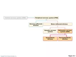
Central nervous system (CNS). Peripheral nervous system (PNS). Sensory (afferent) division. Motor (efferent) division. Somatic nervous system. Autonomic nervous system (ANS). Sympathetic division. Parasympathetic division. Figure 14.1. Neuro- transmitter at effector.

Central nervous system (CNS)
E N D
Presentation Transcript
Central nervous system (CNS) Peripheral nervous system (PNS) Sensory (afferent) division Motor (efferent) division Somatic nervoussystem Autonomic nervous system (ANS) Sympathetic division Parasympathetic division Figure 14.1
Neuro- transmitter at effector Cell bodies in central nervous system Effector organs Peripheral nervous system Effect Single neuron from CNS to effector organs ACh + SOMATIC NERVOUS SYSTEM Stimulatory Heavily myelinated axon Skeletal muscle Two-neuron chain from CNS to effector organs NE ACh Unmyelinated postganglionic axon Ganglion SYMPATHETIC Lightly myelinated preganglionic axons + Epinephrine and norepinephrine ACh Stimulatory or inhibitory, depending on neuro- transmitter and receptors on effector organs AUTONOMIC NERVOUS SYSTEM Adrenal medulla Blood vessel ACh ACh Smooth muscle (e.g., in gut), glands, cardiac muscle PARASYMPATHETIC Lightly myelinated preganglionic axon Unmyelinated postganglionic axon Ganglion Acetylcholine (ACh) Norepinephrine (NE) Figure 14.2
Parasympathetic Sympathetic Eye Eye Brain stem Salivary glands Skin* Cranial Salivary glands Sympathetic ganglia Heart Cervical Lungs Lungs T1 Heart Stomach Thoracic Stomach Pancreas Liver and gall- bladder Pancreas L1 Adrenal gland Liver and gall- bladder Lumbar Bladder Bladder Genitals Genitals Sacral Figure 14.3
Eye Ciliary ganglion CN III Lacrimal gland CN VII Pterygopalatine ganglion Pterygopalatine ganglion Nasal mucosa CN IX CN X Submandibular ganglion Submandibular and sublingual glands Otic ganglion Parotid gland Heart Cardiac and pulmonary plexuses Lung Liver and gallbladder Celiac plexus Stomach Pancreas S2 Large intestine S4 Pelvic splanchnic nerves Small intestine Rectum Inferior hypogastric plexus Urinary bladder and ureters Preganglionic Genitalia (penis, clitoris, and vagina) Postganglionic Cranial nerve Figure 14.4
Sympathetic (Thoracolumbar) Division • Preganglionic neurons are in spinal cord segments T1 – L2 • Sympathetic neurons produce the lateral horns of the spinal cord
Eye Lacrimal gland Nasal mucosa Pons Sympathetic trunk (chain) ganglia Blood vessels; skin (arrector pili muscles and sweat glands) Superior cervical ganglion Salivary glands Middle cervical ganglion Heart Inferior cervical ganglion Cardiac and pulmonary plexuses Lung T1 Greater splanchnic nerve Lesser splanchnic nerve Liver and gallbladder Celiac ganglion L2 Stomach Superior mesenteric ganglion White rami communicantes Spleen Adrenal medulla Kidney Sacral splanchnic nerves Lumbar splanchnic nerves Small intestine Inferior mesenteric ganglion Large intestine Rectum Preganglionic Postganglionic Genitalia (uterus, vagina, and penis) and urinary bladder Figure 14.6
Spinal cord Dorsal root Ventral root Rib Sympathetic trunk ganglion Sympathetic trunk Ventral ramus of spinal nerve Gray ramus communicans White ramus communicans Thoracic splanchnic nerves (a) Location of the sympathetic trunk Figure 14.5a
Stimulus Dorsal root ganglion Sensory receptor in viscera 1 Spinal cord Visceral sensory neuron 2 Integration center • May be preganglionic neuron (as shown) • May be a dorsal horn interneuron • May be within walls of gastrointestinal tract 3 Autonomic ganglion Efferent pathway (two-neuron chain) • Preganglionic neuron • Ganglionic neuron 4 Visceral effector 5 Response Figure 14.7
Referred Pain • Visceral pain afferents travel along the same pathway as somatic pain fibers • Pain stimuli arising in the viscera are perceived as somatic in origin
Heart Lungs and diaphragm Liver Heart Gallbladder Liver Appendix Stomach Pancreas Small intestine Ovaries Colon Kidneys Urinary bladder Ureters Figure 14.8
Neurotransmitters • Cholinergic fibers release ACh • All ANS preganglionic axons • All parasympathetic postganglionic axons • Adrenergic fibers release NE • Most sympathetic postganglionic axons • Exceptions: sympathetic fibers secrete ACh at sweat glands, some blood vessels in skeletal muscles
Two-neuron chain from CNS to effector organs NE ACh Unmyelinated postganglionic axon Ganglion SYMPATHETIC Lightly myelinated preganglionic axons + Epinephrine and norepinephrine ACh Stimulatory or inhibitory, depending on neuro- transmitter and receptors on effector organs AUTONOMIC NERVOUS SYSTEM Adrenal medulla Blood vessel ACh ACh Smooth muscle (e.g., in gut), glands, cardiac muscle PARASYMPATHETIC Lightly myelinated preganglionic axon Unmyelinated postganglionic axon Ganglion Acetylcholine (ACh) Norepinephrine (NE) Figure 14.2
Receptors for Neurotransmitters • Cholinergic receptors for ACh • Adrenergic receptors for NE
Cholinergic Receptors • Two types of receptors bind ACh • Nicotinic • Muscarinic • Named after drugs that bind to them and mimic ACh effects
Nicotinic Receptors • Found on • Motor end plates of skeletal muscle • All ganglionic neurons (sympathetic and parasympathetic) • Hormone-producing cells of adrenal medulla • Effect of ACh at nicotinic receptors is always stimulatory
Muscarinic Receptors • All effector cells stimulated by postganglionic cholinergic fibers • The effect of ACh at muscarinic receptors • Can be inhibitory or excitatory • Depends on receptor type of target organ
Adrenergic Receptors • Two types • Alpha () (subtypes 1, 2) • Beta () (subtypes 1, 2 , 3) • Effects of NE depend on which subclass of receptor on the target organ
Effects of Drugs • Atropine • Anticholinergic; blocks muscarinic receptors • Prevent salivation during surgery, and dilates pupils for exam • Neostigmine • Inhibits acetylcholinesterase • Used to treat myasthenia gravis
Effects of Drugs • OTC for colds, allergies, and nasal congestion • Stimulate -adrenergic receptors • Beta-blockers • Attach to 2 receptors to dilate lung bronchioles in asthmatics
Sympathetic Tone • Sympathetic controls BP at rest • Sympathetic tone (vasomotor tone) • Keeps BV in a continual state of partial constriction • Alpha-blocker drugs interfere with vasomotor fibers and are used to treat hypertension
Parasympathetic Tone • Parasympathetic dominates heart and smooth muscle of digestive and urinary tract organs • Slows heart • Dictates normal activity levels of digestive and urinary tracts • Sympathetic can override effects during stress • Block parasympathetic increase heart rate and block fecal and urinary retention
Unique Roles of the Sympathetic Division • Adrenal medulla, sweat glands, arrectorpili muscles, kidneys, and most BV receive only sympathetic fibers • Sympathetic controls • Thermoregulatory responses to heat • Release reninfrom kidneys • Metabolic effects • Increases metabolic rate • Raises blood glucose • Mobilizes fats for use as fuels
Effects of Sympathetic Activation • Sympathetic is long lasting because NE • Is inactivated more slowly than ACh • NE and epinephrine released into blood and remain until destroyed by liver
Control of ANS Functioning • Hypothalamus—integrative center of ANS • Subconscious input via limbic lobe
Communication at subconscious level Cerebral cortex (frontal lobe) Limbic system (emotional input) Hypothalamus Overall integration of ANS, the boss Brain stem (reticular formation, etc.) Regulation of pupil size, respiration, heart, blood pressure, swallowing, etc. Spinal cord Urination, defecation, erection, and ejaculation reflexes Figure 14.9
Hypothalamic Control • Control may be direct or indirect (through the reticular system) • Centers of the hypothalamus control • Heart activity and blood pressure • Body temperature, water balance, and endocrine activity • Emotional stages (rage, pleasure) and biological drives (hunger, thirst, sex) • Reactions to fear and the “fight-or-flight” system
Developmental Aspects of the ANS • During youth, ANS impairments are usually due to injury • In old age, ANS efficiency declines, partially due to structural changes at preganglionic axon terminals
Developmental Aspects of the ANS • Effects of age on ANS • Constipation • Dry eyes • Frequent eye infections • Orthostatic hypotension • Low BP occurs because aging pressure receptors respond less to changes in BP with changes in body position and because of slowed responses by sympathetic vasoconstrictor centers