Download
1 / 57
570 likes | 679 Views
Stroke, Head Trauma and conciousness. Amy Wood, Haddy Cosh, Vishal Chauhan, Asfand Baig , Stewart O’Conner. Definition. Definition. a syndrome of rapid onset of cerebral deficit (usually focal) Lasting > 24 hours or leading to death and no cause apparent other than a vascular one.

E N D
Stroke, Head Trauma and conciousness Amy Wood, Haddy Cosh, Vishal Chauhan, AsfandBaig, Stewart O’Conner
Definition • a syndrome of rapid onset of cerebral deficit (usually focal) • Lasting > 24 hours or leading to death and no cause apparent other than a vascular one
Stroke Risk Factors • Non Modifiable • Modifiable
Stroke Risk Factors • Non Modifiable • Age • Male • FHx • Race – black/ hispanic • Modifiable • HT • IHD • AF • DM • Hypercholesterolaemia • Smoking • Alcohol
Types • Ischaemia/ embolism causing cerebral infarct – 80% • Intracebral Haemorrhagic – 15%
Causes -Haemorrhagic • Ruptured aneurysm • Trauma (subarachnoid/intracerebral) • Dissection (carotid/vertebral)
Causes - Ischaemic • Cerebral Thrombosis • Cerebral Emboli • Give examples • Lacunar
Symptoms - General • Weakness/Paralysis or numbness on contralateral side • Vertigo/dizziness • Headache • Visual loss/blurred vision • Faintness • Confusion • Speech problems • Difficulty swallowing • Cognitive problems • Memory problems • Consciousness alterations • BUT…DEPENDS ON SITE
Stroke Syndromes • TACS - Total Anterior Circulation Syndrome • PACS - Partial Anterior Circulation Syndrome • LACS - Lacunar Syndrome • POCS - Posterior Circulation Syndrome
Extras - watersheds • Susceptibility to ischaemia: • Systemic BP drop • ACA-MCA occlusion of carotid
TIA • Sudden focal deficit – usually only a few seconds • Presentation very similar to stroke • Amaurosis fugax?? • <24 hours with complete recovery • Issue: after 1 hour ischaemic damage has already occurred • High risk of recurrence and full stroke
Causes- TIA Carotid artery insufficiency – 80% Veterbrobasilar Insufficiency – 20% Circle of Willis – collateral supplies
Management • Assessment/ diagnosis • Location • Subtype • Cause • Acute intervention • Secondary prevention • Reduce risk factors
Assessment: Diagnosis • Clinically usually • FAST • Imaging - <3hrs • CT • Available • Exclude haemorrhage • MRI • If brainstem or cerebellar symptoms
Acute intervention • Admit to Acute Stroke Unit for assessment • Iscahaemic – Thrombolysis rTPA within 3 hrs of symptoms • Haemorragic – emergency surgery
Acute intervention • Antiplatelet drugs (Aspirin 150-300mg) if infarct • Contraindicated if haemorrhage!! • Monitor/prevent complications • Physiological monitoring for first 72 hours to maintain CO and supply to brain • HR, Temperature, BP, O2 sats, Blood sugar, ECG
Complications • Post-stroke pain/thalamic pain • 1 week- 6 months after stroke • Anywhere in spinothalamic system • Contralateral side referral of pain • Burning + sharp • Hyperalgesia & Allodynia • Treat as for neuropathic pain • TCAs
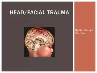
Layers of the brain a) Pia mater b) Arachnoid mater c) Dura mater d) Superior sagittal venous sinus e) Skull f) Falxcelebri g) Subarachnoid space
Pia • Arachnoid • Dura Subarachnoid – arteries Subdural – Bridging veins Epidural – Meningeal arteries
Normal CT • Usually going to be symmetrical • Ventricles symmetrical and equally full
Midline Shift • Coup injury – injury on same side of force • Contra coup– injury on the opposite side on injury • If you see midline shift, you have a high pressure situation
Case 1 • Young lady hit on the side of head by a glass at a gig, seemed to recover , Found slumped 50 minutes later • Ix? • CT/MRI, x-ray if fracture • Where may she have been hit? • Pterion • What bones converge here? • frontal, parietal, sphenoid, temporal • What does this area cover? • Middle meningeal artery • Type of intracranial haemorrhage? • extradural (epi) • Type of blood characterises this? • Arterial • Why passed out? • raised ICP • Rx • surgical
Extraduralhaematoma: • Midline shift • Lenticular shape • This can be middle meningeal artery – pterion bone breaks • Cerebral perfusion pressure = mean arterial pressure – ICP • Extraduralhaematoma you give Mannitol – 100mL at 20% • Diuretic
Case 2 • Old alcoholic man had a fall in the park now noticed to be very drowsy with low consciousness • Ix: • CT/MRI • Likely haematoma? • Subdural • Other symptoms? • Headache, confusion, N/V, tinnitus, speech and visual problems, dizziness, weakness • Where is the bleed likely to be? • bridging veins • Type of blood? • venous • Rx depends on size + growth rate: often conservative (body reabsorbs), sometimes burr-hole drainage • Acute or Chronic
Subdural Haematoma: • Runs along the surface of the brain, underneath the dura • Depending on the GCS score of the patient you may need to remove it • Midline shift
Subarachnoid Haemorrhage • Sudden onset severe headache, often at the back of the head, Neck stiffness, Impaired consciousness (drowsiness / coma), Cranial nerve signs, Hemiplegia • The bleeding occurs as the result of rupture of aneurysm (80%) and AV malformations (15%) or trauma
Contusion (bruise) • Intra- axial • As bruise swells, pressure goes up – all features of raised ICP (coma) • If you remove them you need to do a craniotomy
Diffuse Axonal InjuryRTAs / shaken baby syndrome • If a rotational force is applied, the axons are damaged and you can have damage very far away from the original injury – diffuse axonal injury • Small contusions all over the brain • The worse it looks on the CT scan, the worse the injury in the patient – especially if you see an injury in the brainstem • DAI doesn’t look as bad on CT as some of the other ones, but can be much worse
With a mass lesion why do you not get an immediate loss of consciousness?
Due to an ability to Compensate! Intra cranial vol = vol CSF + vol Brain + vol blood + vol Mass lesion Skull can’t expand Compensation – 10-20 ml CSF in to lumbar cisterns Compensation exceeded Increase in ICP herniation
What are the 3 key symptoms of raised ICP? Papilloedema Headache Nausea and Vomiting