Head Trauma
Head Trauma. Anatomy of Nervous System. The nervous system is composed of Brain Spinal cord The nervous system is divided into: Central nervous system (Brain & Spinal Cord) Peripheral nervous system . Superior view of the skull . Physiology of Nervous System. Cerebral Blood Flow (CBF)


Head Trauma
E N D
Presentation Transcript
Anatomy of Nervous System • The nervous system is composed of • Brain • Spinal cord • The nervous system is divided into: • Central nervous system (Brain & Spinal Cord) • Peripheral nervous system
Physiology of Nervous System • Cerebral Blood Flow (CBF) • Main Arterial Pressure (MAP) • Intracranial Pressure (ICP) • Cerebral Perfusion Pressure (CPP) • CPP = MAP – ICP
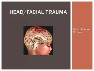
Injuries to the Brain & Skull • Scalp injuries • Skull injuries • Brain injuries
Scalp Injuries • Scalp has many blood vessels so injury may bleed profusely. • Control bleeding with direct pressure. • Don’t apply pressure when there is possible skull injury.
Skull injuries • It include fractures to the cranium and the face, can be associated with brain injury. • It is divided into: • Open skull fracture: cranium is fractures and scalp is lacerated. • Closed skull fracture: scalp is lacerated but cranium is intact. • Basal skull fracture
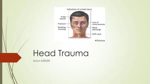
S & S of Skull Fractures and Brain Injuries • Visible bone fragments • Altered mental status • Deep lacerated or severe bruise or hematoma • Depression or deformity of the skull • Severe pain at site of injury • Battle’s Sign • Unequal or unreactive pupils • Raccoon’s eye • Sunken eye • Bleeding from the ears and/or nose • Clear fluid flow from ears and/or nose • Personality change • Increased blood pressure, decreased pulse rate and widening pulse pressure (Cushing’s Syndrome) • Irregular breathing pattern • Temperature increase • Blurred or multiple vision • Impaired hearing or ringing • Equilibrium problems • Forceful or projectile vomiting • Posturing • Paralysis or disability on one side of the body • Seizures • Deteriorating vital signs
Battle’s sign • Indication of fracture of middle cranial fossa of the skull, and may suggest underlying brain trauma. • It appears as a result of extravasation of blood along the path of the posterior auricular artery
Raccoon eyes • Raccoon eyes may be bilateral or unilateral • If unilateral, it is highly suggestive of basilar skull fracture, with a positive predictive value of 85% • Most often associated with fractures of the anterior cranial fossa.
CSF rhinorrhea & otorhea • Suggestive of basal skull fracture
Brain Injuries • Primary (Direct) Brain Injuries • Secondary (Indirect) Injuries
Assessment of TBI • ABC • Alert Verbal Pain Unresponsive • Vital signs • GCS : Eye opening, Best motor response and Best verbal response • History and mechanism of injury
Primary Brain Injuries • It occur at the time of original insult • Direct damage done to brain parenchyma and associated with vascular injuries • Brain tissue can be lacerated, punctured or bruised by broken bones or foreign bodies • Damage is already done • Irreversible • Damage control (debridement)
Secondary Brain Injury • Damage that occurs after the initial insult (ongoing injury processes) • Expanding mass lesions, swelling or bleeding quickly overwhelm buffers • End result is increased intracranial pressure (ICP) and/or herniation • Diagnosis and treatments target minimizing the effects of these indirect insults
Secondary Injury Mechanisms • Mass effect and subsequent elevated ICP and mechanical shifting leading to herniation • Hypoxia • Hypotension and inadequate CBF • Cellular mechanisms
Intracranial Causes • Herniation: displaced brain parenchyma • Damage to brain from trauma against the dura itself as well as producing ischemia as well • Cerebral Edema: intracellular fluid collection within neurons and interstitial spaces. • Intra-cerebral Hematomas
Brain Injuries – Brain Concussion • Usually caused by blunt injuries. • Injuries patient shows transient alteration in neurologic function • Mild injury usually with no detectable brain damage. • May have brief loss of consciousness. • Headache grogginess and short memory loss are common.
Brain Injuries – Brain Contusion • A bruised brain or contusion can occur with closed head injuries. • Usually caused by blow that causes the brain to hit inside the skull • Unconsciousness or decreased level of consciousness can occur
Brain Injuries – A hematoma • Is a collection of blood within tissue. • Hematoma inside the cranium is named according to its location: • Subdural hematoma: blood collection between brain and dura • Epidural hematoma: blood collection between dura and the skull • Subarachnoid Hemorrhage: • Intracerebral hematoma: blood collection within the brain
Epidural Hematomas • Blood between inner table of the skull and the dura • Lens shaped hematomas that do not cross suture lines on CT
Subdural Hematomas • Blood beneath the dura, overlying the brain and arachnoid, resulting from tears to bridging vessels • Crescent shaped density that may run length of skull • Very common in the elderly
Subarachnoid Hemorrhage • Bleeding beneath the arachnoid membrane on the surface of the brain.
Intracranial Hematoma • Focal areas of hemorrhage within the parenchyma
ER Care of Skull Fractures and Brain Injuries • Take appropriate body substance isolation precautions. • Assume spine injury • Monitor conscious patient for changes in breathing • Apply rigid collar, immobilize the neck and spine • Administer high concentration oxygen • Control bleeding • Keep patient at rest • Talk to conscious patient (emotional support) • Dress and bandage open wounds • Mange the patient for shock • Be prepared for vomiting • Transport patient promptly • Monitor vital signs every five minutes