Understanding Prokaryotes: The Diverse Kingdoms of Bacteria and Archaea
460 likes | 568 Views
This overview delves into the fascinating world of prokaryotes, highlighting the two primary domains: Bacteria and Archaea. It explores their structure, differences, and evolution, including insights from Carl Woese's groundbreaking work on rRNA sequences. Prokaryotes are characterized by their simplicity, lack of a nucleus, and unique cellular components such as peptidoglycan layers. The text also covers extremophiles and various adaptations that allow these organisms to thrive in extreme environments, underscoring their role as some of the earliest life forms on Earth.

Understanding Prokaryotes: The Diverse Kingdoms of Bacteria and Archaea
E N D
Presentation Transcript
Justin Ray M. Guce Bio1 2010 SPOTLIGHT: PROKARYOTES(time to shine)
THEN WHAT HAS HAPPENED? • Formation of: (MONERA) • KINGDOM EUBACTERIA • KINGDOM ARCHAEBACTERIA • EUBACTERIA? • ARCHEAEBACTERIA?
THEN WHAT HAS HAPPENED? • Carl Woese (Univ. of Illinois) • Archaea diverged so early in the history of life • rRNA sequence (SSU rRNA, 16s rRNA) • DOMAINS instead of KINGDOM
WHAT is an rRNA • provides a mechanism for decoding mRNA into amino acids • S in 16S represents Svedberg units • S: measures of sedimentation rate
DOMAIN ARCHAEA • DNA exists as a single loop called a plasmid • Outer cell membrane • serves as a barrier • No peptidoglycan • Extremophiles
Examples… • ACIDOPHILES:pH < 5, sulfur springsSulfolobus acidocaldaries S. solfataricus
ALKALIPHILES:-pH > 9, live in soda lakesNatranobacterium gregoryi • HALOPHILES: salt loving, live in Dead Sea & Great Salt LakeHalferax volcanii
THERMOPHILES: heat loving, in acid hot springs, deep ocean geysers • METHANOGENS: CO2 + H2 -->CH4 Methanobacterium • PSYCHROPHILES: cold loving - live in antarctic seas Polaromonas vacuolata
DOMAIN BACTERIA • DNA forms a tangle known as a nucleoid • plasmids, or small loops of DNA • contains PEPTIDOGLYCAN layer
PEPTIDOGLYCAN LAYER • polymer consisting of sugars and amino acids that forms a mesh-like layer outside the plasma membrane • involved in binary fission • Gram-positive bacteria (20 to 80 nm) • Gram-negative bacteria (7 to 8 nm)
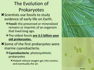
PROKARYOTES • No nucleus, membrane-bound organelles. • Single-celled and unicellular. • The first organism on earth was a very primitive cell similar to bacteria of today. • Immensely successful – they still thrive today. • Very widely distributed.
PROKARYOTES vs. EUKARYOTES
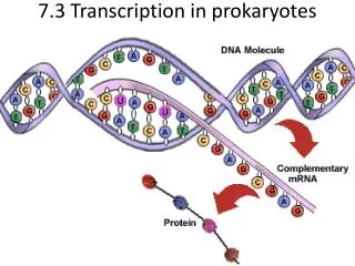
Basic Structures of Prokaryotes • Nucleoid area/region • nucleus-like (“primitive nucleus” = prokaryote) • genetic material (DNA) not surrounded by a nuclear membrane • single circular chromosome
control center of the cell; carries genetic information which directs cellular activities • capable of duplicating itself (replication)
Basic Structures of Prokaryotes • Cytoplasm • Semi-liquid matter which surrounds the nucleoid • consists of water, enzymes, oxygen, waste products, essential nutrients, proteins, carbohydrates and lipids (complex mixture of all the material required by the cell for its metabolic functions)
Basic Structures of Prokaryotes • Ribosomes • sites for protein synthesis • may be found in clusters called polyribosomes • synthesis of proteins rapid due to the proximity of DNA, RNA and ribosomes
Basic Structures of Prokaryotes • Cell membrane • consists of proteins and phospholipids • selectively permeable (controls which substances can enter or leave the cell) • many metabolic reactions take place on the cell membrane enzymes embedded in membrane
Basic Structures of Prokaryotes • Mesosomes • inward folding of the cell membrane • where cellular respiration takes place (also photosynthesis in some)
Basic Structures of Prokaryotes • Cell wall • rigid exterior layer that defines the shape of the cell • main component is peptidoglycan (polysaccharide chains linked together by peptide chains) • thickness of cell wall and exact composition varies
Gram Staining • Gram positive – have many layers of peptidoglycan combined with teichoic acid stains purple/blue • Gram negative – much thinner layer of peptidoglycan, but has an outer layer of lipid (outer membrane) stains red/pink
Basic Structures of Prokaryotes • Capsule • layer of material outside the cell wall • slimy/gelatinous material (complex sugars) produced by the cell membrane • present in some (e.g. Streptococcus penumoniae) • for movement (gliding/sliding)
Basic Structures of Prokaryotes • Flagella • for motility • produces whip-like motion
Basic Structures of Prokaryotes • Pili • hair like tubes with rigid structure • for attachment to other bacteria, tissue; site for viruses to attach • for transfer of genetic material between bacterial cells (conjugation) sex pilus
Basic Structures of Prokaryotes • Spores/Endospores • formed when moisture and nutrient supply is low sporulation • composed of the bacterial DNA wrapped in protein coat
resistant to heat, drying and chemicals • for survival under adverse conditions (not reproduction!) • e.g. Bacillus and Clostridium