Download

1 / 29
290 likes | 397 Views
Designing and Constructing a Set of Fundamental Cell Models: Application to Cardiac Disease. James B.Bassingthwaighte University of Washington Seattle. Engineering and reverse engineering the route from Genome to Function: (Integrating Biological Systems Knowledge). Health. Organism.

E N D
Designing and Constructing a Set of Fundamental Cell Models:Application to Cardiac Disease James B.Bassingthwaighte University of Washington Seattle
Engineering and reverse engineering the route from Genome to Function:(Integrating Biological Systems Knowledge) Health Organism The Physiome Project Organ http://www.physiome.org Tissue • Structure and Function: • Biomedical Problem Formulation • Quantitative Approaches • Engineering Methods • Mechanistic System Modeling • Databasing & Dissemination Cell Molecule Genes
The Physiome and the Physiome Project • The “Physiome”, like the Genome, is a quantitative description of the functional behavior of the physiological state of an individual of a species. In its fullest form it should define relationships from organism to genome and vice versa. • The “Physiome Project” is a concerted effort to define the Physiome through databasing and through the development of a sequence of model types: schema of interactions, descriptions of structure and function, logical prediction, and integrative quantitative modeling for critical projections.
Physiome and Physiome Project • The models of genomic, metabolic, or integrative systems should, via iteration with carefully designed experiments, resolve contradictions amongst prior observations and interpretations. • Reasonably comprehensive and accurate models will demonstrate emergent properties. This is the “reverse engineering” of biology. Some of these will be applied to clinical diagnosis and the evaluation of care. • Databases, concepts, descriptions, and models are to be put in the public domain, an open system.
Structure with Function • The Genome, and the Transcriptome. THE PHYSIOME: • The physico-chemical status. • Descriptions of the Proteome, of solutes, bilayers, organelles, organs, organisms. • Quantitative measures of structural components, e.g. protein and solute levels in cells and organelles, volumes, surface areas, material properties, etc. (The Morphome) • Schema of interactions between the components. Regulatory apparatus for gene expression and metabolism. (The Metabolome) • Computational models (genes +milieu organism).
Three Incentives for Developing the Physiome • To develop understanding of a mechanism or a phenomenom: basic science. • To determine the most effective targets for therapy, either pharmaceutic or genomic. • To design artificial or tissue-engineered, biocompatible implants.
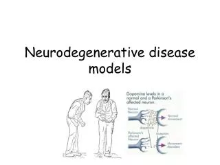
An example: LBBBLeft Bundle Branch Block of the Cardiac Conduction System • Auscultation: Reverse splitting of the second heart sound • ECG: Wide QRS complex and often late T wave • X-ray: Moderate cardiac enlargement • Thallium scan: Low flow in the septum • PET scan: Decreased septal glucose uptake, • but normal septal fatty acid uptake. • The imaging gave three clues to the physiology. • How can the observations be explained?
Electrical activation of the normal heart Prinzen et al., 2000
Schematics of electrical activation RV apex pacing left bundle branch block X Prinzen et al., 2000
Explaining what is observed inLeft Bundle Branch Block • ECG: Wide QRS complex and often late T wave The RBB is activated normally, and excitation proceeds normally over the RV, but since the left branch of the bundle of His is blocked the spread of activation into the left ventricular muscle is delayed 50 to 100 ms, broadening the QRS complex, and delaying the repolarization phase (late T wave)
MRI tagging of Cardiac Contraction Pacing spike ECG ... Presat. pulse 130ms 90ms 50ms ... Gx RF Delay = 50 ms Delay = 90 ms Delay = 130 ms Tagging pulse (Prinzen, Hunter, Zerhouni,1999)
Explaining what is observed inLeft Bundle Branch Block • Auscultation: Reversed splitting of S2 (second heart sound) The RBB is activated normally, but activation of the left ventricle is delayed 50 to 100 ms, so that aortic valve closing is delayed and is later than pulmonic closing, rather than earlier. During inspiration increased RV filling, delaying pulmonic valve closure, shortens (rather than lengthening) the interval between pulmonic and aortic valve closure: reversed respiratory influence on second sound splitting interval. Normally, Insp longer A2–P2 , but here Insp shorter P2–A2
Effect of RV apex pacing on regional LV epicardial fiber strain Segment length early-activated late-activated Prinzen et al, Am. J. Physiol, 1990
Atrial pacing RV apex pacing LV free wall pacing anterior base septum apex posterior Prinzen et al, J Am Coll Cardiol, 1999 Distribution of external work in the LV wall (mJ/g) 8 0 0
To explain what is seen in LBBB: • Thallium scans: Decreased septal blood flow relative to rest of LV because local demand is reduced. Decreased septal mass due to local atrophy. • PET Glucose Uptake: Decreased septal uptake due to shift away from glucose with diminished demand relative to supply. PET data show normal FA uptake. Regional FA uptake is matched to local flow. • X-ray: LV hypertrophy: Hypertrophic free wall due to increased workload and low contractile efficiency. This is partially attributable to increased wall tension with LV cavity volume increase: T=PxR.
Cardiac fiber structuring: LV base LV near the apex From Torrent-Guasp, 1998
Rabbit Heart: Epicardial fibers – blue Subendocardial fibers - yellow From Vetter and McCulloch, UCSD
Integration by Computation: The Cardiome • Transport: • UW: Flows, uptake (O2, fats) • Cardiac Mechanics: • Auckland Univ: P.Hunter • UCSD: McCulloch • Maastricht: Arts, Prinzen, Reneman • JHU: W.Hunter • Action Potentials: • Oxford U: D. Noble • Johns Hopkins: Winslow • Case-Western: Rudy • Cardiac excitatory spread: • CWRU: Rudy et al. • Johns Hopkins: Winslow • Syracuse: Jalife • UCSD: McCulloch N.Smith, P. Hunter,et al. 1998
What are the mechanisms for the responses in Left Bundle Branch Block? • Thallium scans: How is local flow regulated? • PET Glucose Uptake: How is glycolysis regulated? • MR Strain Patterns: How do structure, excitation, and contraction combine to produce these? • X-ray LV hypertrophy: What regulates actin and myosin expression?
Excitation-Contraction Coupling • Mechanical Feedback • Cooperativity
WEAK ATP ADP • Mechanical Feedback Energy Use depends on shortening velocity: g +g V 1 0 ADP ATP f STRONG • Weak-Strong vs. Attached-Detached Landesberg/Sideman, 1998
The Conservative Phys-chem cell(no protein synthesis or proteolyis) • Balances mass, charge, volume, energy, reducing equivalents, concentrations • Serves as a primitive for expanded models • RBC, prokaryote, eukaryote, myocyte, B-cell • Serve as entry to databases • Component of healthy and diseased tissues • Basis for multicellular integrated systems models • Understanding via metabolic control analysis of networks • Test bed for mechanistic pharmacodynamic models and selection for drug design and for genomic intervention
The glycolytic conservative cell (with eternal proteins) Ca2+ e.g. arep.med.harvard.edu, Edwards, Palsson,Church et al. 3Na+ Substrates ATP Ca2+ Ca2+ Metabolites K+ Glycolysis Charge neutrality H+ pH balance ~P balance Purine balance Osmotic balance Water balance Na+ RBC Redox state Free Energy calmodulin Na+ 3Na+ ATP 2K+ H+
The conservative cell with eternal proteins INaCa Ip(Ca) Substrates ATP Ca2+ Ca2+ Na+ Glycolysis, fatty acid K+ Charge neutrality H+ TCA OxPhosph pH ~P balance Purine balance Osmotic balance Water balance Redox state Free Energy calmodulin Endoplasmic reticulum ATP Na+ Na+ Na+ ATP K+ H+ INaK INa
The sustainable metabolic muscle cell OxPhosph INaCa ICa,b Ip(Ca) ICa,K Substrates TCA ATP Ca2+ Ca2+ Ca2+ Na+ K+ ICa T-tubule K+ IKr K+ subspace Ca2+ IKs pH, P & Charge neutrality K+ RyR IK1 calseq K+ ATP regulation IKp Ca2+ calmodulin Ca2+ calmodulin K+ TRPN Ca2+ Leak Ito1 Sarcoplasmic reticulum ATP Na+ Ca2+ Na+ Na+ Na+ ATP K+ H+ INaK INa INa,b
The cardiac muscle cell INaCa ICa,b Ip(Ca) ICa,K Substrates ATP Ca2+ Ca2+ Ca2+ Na+ K+ ICa T-tubule K+ Glycolysis IKr K+ subspace OxPhosph Ca2+ IKs TCA K+ RyR IK1 calseq K+ IKp Ca2+ calmodulin Ca2+ K+ Leak Ito1 Sarcoplasmic reticulum ATP Na+ Ca2+ Na+ Na+ Na+ ATP (building from Luo-Rudy 1994-2001 and Winslow et al. 1999) K+ H+ INaK INa INa,b
Conclusions: • Conservative cell models provide a basis for a host of specific applications. • Their behavior is innately complex and highly dependent on the conditions. • Computability is a major issue if models are to be used are practical aids to thinking. • Even now they provide short-term prediction of the consequences of intervention.