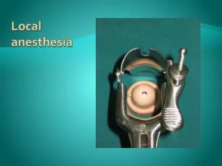

Local Anesthesia
E N D
Presentation Transcript
LECTURE 2Localanesthesia. Indicationsandcontraindications. Technique. Complicationsoflocalanesthesia, theirtreatmentandprophylaxis. Removaloftheteeth: instruments, indicationsandcontraindications, patientpreparation, complicationsduringtoothextractionandinthepostoperativeperiod, theirtreatmentandprophylaxis.
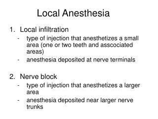
Local Anesthesia Local infiltration type of injection that anesthetizes a small area (one or two teeth and asscociated areas) anesthesia deposited at nerve terminals Nerve block type of injection that anesthetizes a larger area anesthesia deposited near larger nerve trunks
Local anaesthesia • Methods: • Reducing temperature. • Is used only to produce surface anaesthesia e.g. ethyl chloride spray. • Physical damage to nerve trunk e.g. nerve sectioning. • Unsafe for therapeutic uses, only in Trigeminal Neuralgia. • Chemical damage to nerve trunk e.g. neurolytic agents. • Silver nitrate, Phenol - Unsafe for therapeutic use.
Local anaesthesia • Methods:Cont • Anoxia or hypoxia resulting in lack of oxygen to nerve. • Unsafe as well. • Stimulation of large nerve fibres, blocking the perception of smaller diameter fibres. • includes Acupuncture and TENS (Transcutaneous Electronic Nerve Stimulation) • Drugs that block transmission at sensory nerve endings or along nerve fibres. • There action is fully reversible and without permanent damage to the tissues.
Classification: • Classified according to their chemical structures and the determining factor is the intermediate chain, into two groups: Ester Amide • They differ in two important respect: • Their ability to induce hypersensitivity reaction. • Their pharmacokinetics - fate and metabolism.
Types of Nerve Anesthesia • Maxillary • posterior superior alveolar block • middle superior alveolar block • anterior superior alveolar block • greater palatine block • infraorbital block • nasopalatine block • Mandibular • inferior alveolar block • buccal block • mental block • incisive block • Gow-Gates mandibular nerve block
Considerations • dental procedures can usually commence after 3 – 5 minutes • failure requires re-administration using another method • never re-administer using the same method • keep in mind the total # of injections and the dosages • never inject into an area with an abcess, or other type of abnormality
Maxillary Nerve Anesthesia • Chart 9-1 • pulpal anesthesia: through anesthesia of each nerve’s dental branches as they extend into the pulp tissue (via the apical foramen) • periodontal: through the interdental and interradicular branches • palatal: soft and hard tissues of the palatal periodontium (e.g. gingiva, periodontal ligaments, alveolar bone) • PSA block: recommended for maxillary molar teeth and associated buccal tissues in ONE quadrant • MSA block: recommended for maxillary premolars and associated buccal tissues • ASA block: recommended for maxillary canine and the incisors in ONE quadrant • greater palatine block: recommended for palatal tissues distal to the maxillary canine in ONE quadrant • nasopalatine block: recommended for palatal tissues between the right and left maxillary canines
PSA Nerve Block • figures 9-2 through 9-7 • pulpal anesthesia of the maxillary 3rd, 2nd and 1st molars • required for procedures involving two or more molars • sometimes anesthesia of the 1st molar also required block of the MSA nerve • associated buccal periodonteum overlying these molars • including the associated buccal gingiva, periodontal ligament and alveolar bone • useful for periodontal work on this area
PSA Nerve Block • target: PSA nerve • as it enters the maxillar through the PSA foramen on the maxilla’s infratemporal service – Figure 9-2 & 9-3 • into the tissues of the mucobuccal fold at the apex of the 2nd maxillary molar (figures 9-4 and 9-5) • mandible is extended toward the side of the injection, pull the tissues at the injection site until taut • needle is inserted distal and medial to the tooth and maxilla • depth varies from 10 to 16 mm depending on age of patient • no overt symptoms (e.g. no lip or tongue involvement) • can damage the pterygoid plexus and maxillary artery
MSA Nerve Block • limited clinical usefulness • can be used to extend the infraorbital block distal to the maxillary canine • can be indicated for work on maxillary pre-molars and mesiobuccal root of 1st molar (Figure 9-8) • if the MSA is absent – area is innervated by the ASA • blocks the pulp tissue of the 1st and 2nd maxillary premolars and possibly the 1st molar + associated buccal tissues and alveolar bone • useful for periodontal work in this area • to block the palatine tissues in this area – may require a greater palatine block
MSA Block • target area: MSA nerve at the apex of the maxillary 2nd premolar (figures 9-8 and 9-9) • mandible extended towards injection site • stretch the upper lip to tighten the injection site • needle is inserted into the mucobuccal fold • tip is located well above the apex of the 2nd premolar • figure 9-11 • harmless tingling or numbness of the upper lip • overinsertion is rare
ASA Block • figures 9-12 through 9-14 • can be considered a local infiltration • used in conjunction with an MSA block • the ASA nerve can cross the midline of the maxilla onto the opposite side! • used in procedures involving the maxillary canines and incisors and their associated facial tissues • pulpal and facial tissues involved – restorative and periodontal work • blocks the pulp tissue + the gingiva, periodontal ligaments and alveolar bone in that area
ASA Block • target: ASA nerve at the apex of the maxillary canine – figures 9-12 & 9-13 • at the mucobuccal fold at the apex of the maxillary canine – figure 9-13 • harmless tingling or numbness of the upper lip • overinsertion is rare
Infraorbital Nerve Block • figures 9-15 through 9-17 • anesthetizes both the MSA and ASA • used for anesthesia of the maxillary premolars, canine and incisors • indicated when more than one premolar or anterior teeth • pulpal tissues – for restorative work • facial tissues – for periodontal work • also numbs the gingiva, periodontal ligaments and alveolar bone in that area • the maxillary central incisor may also be innervated by the nasopalatine nerve branches
IO Block • target: union of the ASA and MSA with the IO nerve after the IO enters the IO foramen – figure 9-15 • also anesthesizes the lower eyelid, side of nose and upper lip • IO foramen is gently palpated along the IO rim • move slightly down about 10mm until you feel the depression of the IO foramen – figure 9-16 • locate the tissues at the mucobuccal fold at the apex of the 1st premolar • place one finger at the IO foramen and the other on the injection site – figure 9-17 • locate the IO foramen, retract the upper lip and pull the tissues taut • the needle is inserted parallel to the long axis of the tooth to avoid hitting the bone • harmless tingling or numbness of the upper lip, side of nose and eyelid
Greater Palatine Block • figures 9-19 through 9-21 • used in restorative procedures that involve more than two maxillary posterior teeth or palatal tissues distal to the canine • also used in periodontal work – since it blocks the associated lingual tissues • anesthetizes the posterior portion of the hard palate – from the 1st premolar to the molars and medially to the palate midline • does NOT provide pulpal anesthesia – may also need to use ASA, PSA, MSA or IO blocks • may also need to be combined with nasopalatine block
Greater Palatine Block • target: GP nerve as it enters the GP foramen • located at the junction of the maxillary alveolar process and the hard palate – at the maxillary 2nd or 3rd molar – figure 9-19 • palpate the GP foramen – midway between the median palatine raphe and lingual gingival margin of the molar tooth – figure 9-21 • can reduce discomfort by applying pressure to the site before and during the injection • produces a dull ache to block pain impulses • also slow deposition of anesthesia will also help • needle is inserted at a 90 degree angle to the palate – figure 9-22
Nasopalatine Block • figure 9-23 through 9-26 • useful for anesthesia of the bilateral portion of the hard palate • from the mesial of the right maxillary 1st premolar to the mesial of the left 1st premolar • for palatal soft tissue anesthesia • periodontal treatment • required for two or more anterior maxillary teeth • for restorative procedures or extraction of the anterior maxillary teeth – may need an ASA or MSA block also • blocks both right and left nerves
Nasopalatine Block • target: both right and left nerves as they enter the incisive foramen from the mucosa of the anterior hard palate – figure 9-23 & 9-25 • posterior to the incisive papilla • injection site is lateral to the incisive papilla – figure 9-26 • head turned to the left or right • inserted at a 45 degree angle about 6-10 mm – gently contact the maxillary bone and withdraw about 1mm before administering • can reduce discomfort by applying pressure to the site before and during the injection • produces a dull ache to block pain impulses • also slow deposition of anesthesia will also help • can anesthetize the labial tissues between the central incisors prior to palatal block • can block some branches of the nasopalatine prior to injection
Mandibular Blocks • Chart 9-2 • infiltration is not as successful as maxillary anesthesia • substantial variability in the anatomy of landmarks when compared to the maxilla • pulpal anesthesia: block of each nerve’s dental branches • periodontal: through the interdental and interradicular branches • Inferior Alveolar block: for mandibular teeth + associated lingual tissues and for the facial tissues anterior to the mandibular 1st molar • Buccal block: tissues buccal to the mandibular molars • Mental block: facial tissues anterior to the mental foramen (mandibular premolars and anterior teeth) • Incisive block: for teeth and facial tissue anterior to the mental foramen • Gow-Gates: most of the mandibular nerve • for quadrant dentistry
Inferior Alveolar Block • also called the mandibular block • most commonly used in dentistry • for restorative, extraction and periodontal work • pulpal anesthesia for extractions and restorative • lingual periodonteal anesthesia • facial periodonteal anesthesia of anterior mandibular teeth and premolars • may be combined with the buccal block • can overlap with the incisive block • local infiltrations in the anterior area are more successful than posterior injections • variability in the location of the mandibular foramen on the ramus can lessen the success of this injection • usually avoid bi-lateral injections since they will completely anesthetize the entire tongue and can affect swallowing and speech
IA Block • target: slightly superior to the mandibular foramen – figure 9-27 • the medial border of the ramus • will also anesthetize the adjacent anterior lingual nerve – figure 9-30 • injection site is found using hard landmarks • palpate the coronoid notch – above the 3rd molar • imagine a horizontal line from the coronoid notch to the pterygomandibular fold which covers the pterygomandibular raphe – figure 9-32 • this fold becomes more prominent as the patient opens their mouth wider • refer to video notes • figure 9-33 • needle is inserted into the pterygomandibular space until the mandible is felt – retract about 1 mm • average depth: 20-25mm • diffusion of anesthesia will affect the lingual nerve
IA block • symptoms: harmless tingling and numbness of the lower lip due to block of the mental nerve • tingling and numbness of the body of the tongue and floor of mouth – lingual nerve involvement • complications: • failure to penetrate enough can numb the tongue but not block sufficiently • lingual shock – involuntary movement as the needle passes the lingual nerve • transient facial paralysis – facial nerve involvement if inserted into the deeper parotid gland – figure 9-34 • inability to close the eye and drooping of the lips on the affected side • hematoma can occur • some muscle soreness • patient-inflicted trauma – lip biting etc...
Buccal Block • figures 9-36 and 9-37 • for buccal periodonteum of mandibular molars, gingiva, periodontal ligament and alveolar bone • for restorative and periodontal work • buccal nerve is readily located on the surface of the tissue and not within bone
Buccal block • target: buccal nerve as it passes over the anterior border of the ramus through the buccinator – figure 9-36 • injection site is the buccal tissues distal and buccal to the most distal molar – on the anterior border of the ramus as it meets the body – figure 9-37 • pull the buccal tissue tight and advance the needle until you feel bone – only about 1 to 2mm figure 9-38 • patient-inflicted trauma – lip biting etc...
Mental Block • figures 9-39 through 9-41 • for facial periodonteum of mandibular premolars and anterior teeth on one side • for restorative work – incisive block should be considered instead
Mental Block • target site: mental nerve before it enters the mental foramen where it joins with the incisive nerve to form the IA nerve – figure 9-39 • palpate the foramen between the apices of the 1st and 2nd premolars • palpate it intraorally – find the mucobuccal fold between the apices of the 1st and 2nd premolars – figure 9-42 • in adults, the foramen faces posterosuperiorly • may be anterior or posterior • can be found using radiographs • insertion site is the mucobuccal fold tissue directly over or slight anterior to the foramen site • avoid contact with the mandible with the needle • depth is 5 to 6mm • no need to enter the foramen
Incisive Block • for pulp and facial tissues of the teeth anterior to the mental foramen • same as the mental block except pulpal anesthesia is provided also • restorative and periodontal work • IA block indicated for extractions – no lingual anesthesia with an incisive block • target: mental foramen – figure 9-43
Incisive Block • injection site: figure 9-44 • same as for the mental block • directly over or anterior to the mental foramen • in the mucobuccal fold at the apices of the 1st and 2nd premolars • pull the buccal tissues laterally • more anesthesia is used for this block when compared to the mental block • pressure is applied during the injection – forces for anesthetic solution into the foramen and block the deeper incisive nerve • the increased injection solution may balloon the facial tissues
Gow-Gates • figures 9-45 through 9-50 • blocks the IA, mental, incisive, lingual, mylohyoid, auriculotemporal and buccal nerves – figure 9-28 and 9-45 • used for quadrant dentistry • buccal and lingual soft tissue from most distal molar to the midline • greater success than an IA block
Gow-Gates • target site: anteromedial border of the mandibular condylar neck – figure 9-46 • just inferior to the insertion of the lateral pterygoid muscle • injection site is intraoral • locate the intertragic notch and labial commisure extraorally • draw a line from the tragus/intertragic notch to the labial commisure – figure 9-47 • place your thumb on the condyle (just in front of the tragus when the mouth is open) • pull buccal tissue away • place the needle inferior to the mesiolingual cusp of the MAXILLARY 2nd molar • the needle penetrates distal to the maxillary 2nd molar • see the video
Indications and contra-indications to removal of permanent teeth. Indications to planned tooth removal: • 1.)Unsuccessfulness of endodonthyc treatment with presence of the chronic inflammation of periodontium and adjoining tissues of a bone. This intervention is especially indicated in case of chronic intoxications of the patient with odontogenic intoxication centres (chroniosepsis) • 2.) Impossibility of conservative treatment through considerable crown destruction or the technical obstacles connected with anatomic features, treatment errors, caused by root perforation.
3.) Total destruction of crown part of the tooth, impossibility of using the root for tooth prosthetics. • 4.) Mobility of ІІІ degree and tooth promotions as a result of resorption of bone round a cell with presence of heavy forms of a periodontosis and parodontitis. • 5.) Atypically placed teeth which injure a mouth mucous membrane, tongue, and which can't be treated by ortodonthic treatment. • 6.) Unteethed in time or partially teethed teeth which predetermine inflammatory processes in adjoining tissues, which cannot be liquidated some other way.
7.) Placed in crisis cracks, teeth do impossible reposition of fragments andcan't be treated by conservative treatment. • 8.) Outstanding as a result of loss of the antagonist teeth, teeth which convergence and divergence, disturb embarrass the process of manufacturing tooth prosthetics. treatment. For elimination of anomalies of a bite (occlusion) during the orthodontic treatment, intact teeth removal is also indicated.
Contra-indications. A number of inflammatory and local diseases, and also some physiologic conditions are contra-indications to this intervention. Removal of tooth at such patients can be done after preparation and treatment.
Relative contra-indications to operations of tooth removal are: • 1.) Cardiovascular diseases (preinfarction conditions and 3-6 month after the infarction of a myocardium, hypertonic illness in crisis. IHD(ischemic heart disease), paroxysm, blinking arhythmia, paroxysmal tachycardia, acute septic endocarditis); • 2.) Acute diseases of parenchymatosic organs - liver, kidneys, pancreas (an infectious hepatitis, (glomerunonephritis); • 3.) Haemorragical diseases (a hemophilia, illness of Verlgof, agranulocytosis, acute leukemia); • 4.) acute infectious diseases (a flu, ARVD(acute respiratoric virus disease), a pneumonia);
5.) disease of CNS (central neuronic system), (acute disorder of encephal blood circulation, a meningitis); • 6.) Mental (psychological) diseases in an aggravation period (a schizophrenia, a psychosis, an epilepsy); • 7.) acute radiation sickness І - ІІІ degrees; • 8.) disease of a mucous membrane of a mouth (a stomatitis, gingivitis, cheilitis).
Preparation of tooth removal: • -Inspection. • -Preparation of the patient. • -Preparation of doctor’s hands. • -Preparation of the operation field.
Technique of tooth removal: • Tooth removal consists in violent rupture of tissues which connect root with walls of a cell and gums, and its deducing from a cell. During removal of the distorted roots from a cell, its walls are being replaced and the entrance to it extends. Tooth removal is being made by special tools, forceps and elevators. In certain cases tooth extraction by using this tool is impossible. Then a drill for bone removal is used. (operation of root cutting)
Forceps and elevetaors for teeth removal: • Forceps. Under the process teeth removal a lever principle is used. Forceps consists of: cheeks, handles and the lock. In some kind’s of forceps between cheeks and the lock there is a transitive part. Cheeks are used to cover the root or a crown. The handle – a part which is used to hold the forceps. The lock is placed between the handle and a cheek. • For the best fixing of tooth or a root, cheeks have fillets with longitudinal cutting from the inside. The external surface of handles on significant length is relief, internal - smooth. The form of forceps is not the same. Construction depends on anatomical structure of the tooth and it’s place in row of teeth.