Download
1 / 36
430 likes | 1.59k Views
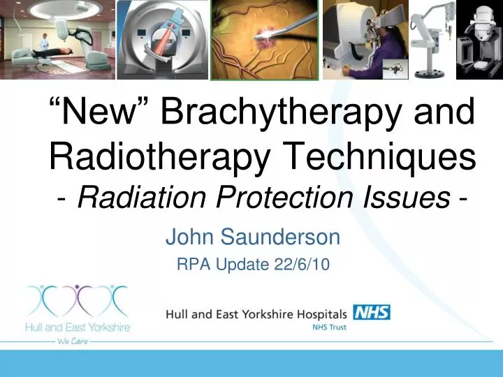
“New” Brachytherapy and Radiotherapy Techniques - Radiation Protection Issues -. John Saunderson RPA Update 22/6/10. Epiretinal Brachytherapy. The Patient’s Problem - Wet AMD. Normal retina. Wet Age Related Macular Degeneration. optic nerve (white disc). Macula lutea (dark area).

E N D
“New” Brachytherapy and Radiotherapy Techniques- Radiation Protection Issues - John Saunderson RPA Update 22/6/10
The Patient’s Problem - Wet AMD Normal retina Wet Age Related Macular Degeneration optic nerve (white disc) Macula lutea (dark area) Abnormal blood vessel growth blood leaking under retina
Current Solution - “Lucentis” 1-3 monthly injections @ £761 per injection “New” Solution - ionising radiation? • 9 Gy will prevent cell division in blood vessels • Retina can tolerate doses at least 25 Gy (in 2Gy fractions)
NOW - Epiretinal beta radiation therapy • 24 Gy to centre of lesion • Single fraction delivered in 3-4 minutes
MERLOT Trial Vidion System, by NeoVista Sr-90 / Y-90 source - 0.5 MeV / 2.3 MeV betas 370 - 555 MBq (Sr-90 29 y half-life) 2.5 mm length x 0.52mm diameter.
Disposable Delivery Module DA Shell Bayonet Shutter Pin Cannula Tip Reusable Delivery Module Cannula Source Positioning Cross Mark Detent Pins
For 24 Gy centre 4Gy @ 4mm radius
Radiation Protection Issues (i) • RSA93/EPR10 • HDR but not HASS • Category 5 registration sufficient • Stored at Eye Hospital - theatre manager source custodian • M(ARS) • consultant oncologist obtained research ARSAC license.
IRMER • Procedures written by Radiation Physics & Theatre Manager and ratified by Ophthalmic Business Meeting • Referrer - ophthalmic surgeon • Practitioner - ARSAC license holder (oncologist) • Operators - ophthalmic surgeon, nurse practitioner, physicists (other theatre staff in future) • Adequate training from NeoVista & in-house • CDG2009 • Reusable type A package
IRR99 - The RPA bits • 2.3 MeV beta range • 1 cm water • 10 m air • (Will be less after attenuation by source capsule and cannula) • Bremßtrahlung • from capsule & cannula • from shielding
Beta particle dose • Vitreous humour acts as beta shield • Source deployed – max. finger dose • @ 4cm in air 1,400. mSv/h (0.4 mGy/s) • @ 4cm source in eye 0.7 mSv/h Very low risk from normal use (4cm is closest point of operator’s finger from deployed source)
Ready to deploy 1070 620 792 250 1800 750 410 40 12 1560 microsieverts per hour Courtesy of Rainer Pintaske & NeoVista
microsieverts per hour #7 Not deployed 6 Sv/h @ 30 cm 0.8 Sv/h @ 100 cm #2 #7 Fully deployed in eye 24 Sv/h @ 30 cm 2.5 Sv/h @ 100 cm #2 microsieverts per hour Courtesy of Rainer Pintaske & NeoVista
Typical Finger dose Scenario • Source ready to deploy • 792 Sv/h x holding 1 minute • 13 Sv • Transit dose from deploying • 100 Sv • Treatment dose • 670 Sv/h x 3.5 min • 39 Sv • Transit dose from deploying • 100 Sv • Cannula removed and end • 792 Sv/h x holding 1 min. • 13 Sv Total finger dose • 265Sv / patient • 150mSv 566 patients
Physicist • 0.038 mSv fingertips • 0.006 mSv to body Effective dose
Effective dose • Highest predicted dose - Medical Physicist • 6 Sv per patient • If 566 patients/y 3.4 mSv/y i.e. surgeon finger dose is critical value • Our measurements, 4 patients • Surgeon’s finger = 0.15 mSv • (Predicted 1.04 mSv) • Physicist body = 0.01 mSv • (Predicted 0.024 mSv)
Controlled Area, Local Rules, RPS? “Controlled area” - IRR99 Reg 16(1) • special procedures necessary to prevent significant exposure • likely to receive E > 6 mSv in a year (Heye>45mSv/y, Hskin>150mSv/y) • Risk assessment shows b) not likely unless >566 patients per year • (20 in trial)
Special procedures necessary ... ACOP para 248 & 249 elaborated on a)
dose rate >7.5 Sv/h over 1 minute and untrained staff • Undeployed = 6 Sv/h @30cm • Deployed in eye = 24 Sv/h @30cm, 2.5 Sv/h @1m • exclusion or supervision of untrained staff necessary • Physicist, surgeon, nurse practitioner trained in procedure • Some other theatre staff (not anaesthetist yet!) • Controlled area (+ local rules + RPS) ? • Maybe ... maybe not
Accuray Cyberknife • 6 MVx unflattened beam • 5 to 60 mm diameter field at 80cm from focus • No fixed isocentre • 50-200 fields per treatment Conventional linac
Conventional linac - +/-1800 rotation around isocentre - “donut” primary shielding OK • CyberKnife linac - 2.5 sr beam - primary shielding in (nearly) all directions required
add 28 cm add 53 cm add 23 cm Hull & East Yorks. CL1 & CL2 add 13 cm concrete (or 3.7cm steel) add 103 cm? (Note: CyberKnife can only fire up at maximum of 220 to the horizontal)
All barriers primary shielding, except • Ceiling - max. 220 to the horizontal • can be programmed to limit beam directions • Many small fields small USE FACTOR of 0.05 recommended
= fraction of a primary-beam workload directed toward a given primary barrier. Use Factor • Linac - IPEM 75 • floor U=1, walls & ceiling U=0.25 • floor 0.42-0.58, ceiling 0.12, 0.2-0.23 walls • NCRP 151 • Cyberknife, U = 0.05 • Determined by analysis by James Rodgers of 324 treatment sessions at Georgetown University Hospital
Secondary Shielding • leakage predominates • “IMRT”, so each beam on for a longer time • “modulation factor” should be used
IMRT Factor CI = (MU for 1Gy prescribed dose by IMRT) CI = (MU for 1 Gy at 10cm depth at isocentre) Modulation Factor, CK CK = average MU per cGy delivered over all SAD’s and tumour depths • High value, more beam on time per treatment • No effect on primary or patient scatter barrier calculation • Effects leakage shielding • Linac IMRT, CI 2 to 10+ • Cyberknife, CK = 15 recommended
Helical Tomotherapy • vs traditional linac treatment • Unfiltered 6 MV X-ray beam • Slit beam (max 5 cm x 40 cm at 85cm isocentre) • 13 cm lead beam stop (Hi-Art II) • Long beam on time more leakage
Zeiss Intrabeam • 50kV, 0.05mA, Au target
Intrabeam Radiation ProtectionParry, Sutton, Mackay et al (Dundee) • No shielding - 10 mSv/h @ 1m • Procedures take between 15 and 40 minutes. • 2mm lead screens - 40 Sv/h @ 1m • Sufficient for operator & anaesthetist • “local shielding at the treatment site is essential in order to reduce the dose rate in adjacent areas to acceptable levels.”
Intraoperative Electron radiation therapy • IntraOp Mobetron • Mobile electron beam linac • Up to 12 MeV e-’s (6-7 cm range) • IntraOp -“No room shielding is required” • Jodi Davies et al 2001 - Depends on workload - scatter & leakage up to 18 Gy/1000Mu @ 2 m (more towards floor)
KV stereotactic radiosurgery Eric Chell et al, 2009 • iRay system (Oraya Therapeutics Inc) • 100kV, 150mm focus-retina, 3 beams, • 16-24 Gy, 4mm diameter target (90%),
Some References & Useful Documents • CyberKnife • NCRP Report No. 151 Structural Shielding Design & Evaluation for MV X- & Gamma-Ray Radiotherapy Facilities, 2005 • Accuray Site Planning documents - www.accuray.com/Services/Site_Planning.aspx • CyberKnife Shielding Design, Jim Rodger, AAPM Summer School 2007 www.aapm.org/meetings/07ss/documents/RodgersCyberKnifeShieldingpostmeeting.pdf • Tomotherapy • Helical tomotherapy shielding calculation for an existing LINAC treatment room: sample calculation and cautions, Chuan Wu, Fanqing Guo and James A Purdy, Phys. Med. Biol. 51 (2006) N389–N392 • Intraoperative Radiotherapy (IORT) • J. Parry, D. Sutton, C. Mackay, J. O'Neill, S. Eljamei, A. Thompson and A. Munro. Radiation protection aspects of setting up an intra-operative radiotherapy facility. Radiotherapy and Oncology, Volume 76, Supplement 2, September 2005, Pages S202-S203 • Joseph DJ, Bydder S, Jackson L, Corica T, Hastrich D, Oliver D, Minchin D, Haworth A, & Saunders C. Prospective Trial Of Intraoperative Radiation Treatment For Breast Cancer. ANZ J. Surg. 2004; 74: 1043–1048 • Daves JL & Mills MD. Shielding assessment of a mobile electron accelerator for intraoperative radiotherapy. J Applied Clin Med Phys, Volume 2, Number 3, Summer 2001 • Kilovoltage stereotactic radiosurgery • Chell E, Firpo M, Shusterman EM, Hansen S, Gertner M. A Novel Stereotactic Radiosurgical Device for the Treatment of Age-Related Macular Degeneration (AMD). AAPM 2009 • see www.orayainc.com/articles.asp for this and papers