Download
1 / 60
630 likes | 1.22k Views
Respiratory Assessment. Thoracic cage bony conical shape with narrow at top Defined by sternum, 12 pairs ribs and 12 thoracic vertebrae Rib 1-7 attached to sternum via costal cartilages Rib 8,9,10 attached to costal cartilage Rib 11,12 “floating” with free palpable tips

E N D
Respiratory Assessment • Thoracic cage bony conical shape with narrow at top • Defined by sternum, 12 pairs ribs and 12 thoracic vertebrae • Rib 1-7 attached to sternum via costal cartilages • Rib 8,9,10 attached to costal cartilage • Rib 11,12 “floating” with free palpable tips • Anterior thoracic Landmarks; • Suprasternal notch, “U” shaped
Sternum-”breastbone” has three parts: The body The xiphoid The Manubrium Manubriosternal Angle- “Angle of Louis”-is articulation of the manubrium and the body of the sternum; continuous with the 2ed rib; marks site of the tracheal bifurcation into the R & L bronchi Costal Angle- R & L costal margins form angle where meet at xiphoid process
Posterior thoracic Landmarks • Vertebra Prominens • Spinous Processes • Interior Boarder of Scapula • 12th Rib • Anterior Chest • Midsternal Line • Midclavicular Line- bisects the center of each clavicle at point halfway between the palpated sternoclavicular and acromioclavicular joint, near nipple line
Posterior Chest • vertebral Line- midspinal • Scapular line- extends through inferior angle of scapula • Lateral Chest- Lift arms 90* & divide by 3 lines • Anterior axillary- down from anterior axillary fold to where the pectoralis major muscle inserts • Posterior anillary down from posterior axillary fold to where latissimus dorsi muscle inserts
; • Midaxillary line-down from apex of axilla, lies between and parallel to other two • Thoracic cavity • Mediastinum- middle section of thoracic cavity contains esophagus, trachea, heart and great vessels; and the Left & Right pleural cavities • Lung Borders- anterior • Apex 9 highest point of lungs 3-4 cm above inner 3rd of clavicles • Base- lower border, rest on diaphragm about 6th rib in midclavicular line
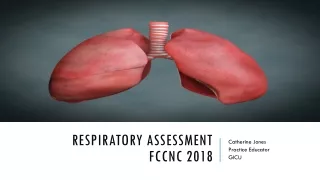
Lungs Border- posterior • C7-Apex of lung tissue • Lungs- 2 pair • Anterior • Right lung shorter because of underlying liver; has 3 lobes • Anterior Right Upper Lobe (RUL) • Right Middle Lobe (RML) • Right Lower Lobe (RLL) - Left Lung- narrower because heart bulges to left; has 2 lobes
Anterior Left Upper Lobe (LUL) • Anterior Left Lower Lobe (LLL) • Posterior • Right Upper Lobe (RUL) and Left Upper Lobe (LUL)- from apices at T1 down to T3 • Right Lower Lobe (RLL) and Left Lower Lobe (LLL)- from the above border to T10 on expiration and to T12 on inspiration. • Pleurae- thin slippery which forms an envelope between lungs and chest wall. • Visceral Pleura- lines outside of lungs down into fissures
Parietal Pleura- lining inside the chest wall and diaphragm. • Trachea- lies anterior to esophagus; is 10– 11 cm long in adult; starts at cricoid cartilage bifurcates below sternal angle into R and L bronchi; posterior bifurcates at T4 or T5; R bronchus shorter wider; L bronchus vertical than L main
Function of Trachea and bronchi- transport gases between environment and lung parenchyma. • Bronchial tree- protect alveoli from small particulate matter in the inhaled air • Bronchi lined- goblet cells which secrete mucus that entraps particles • Bronchi line with cilia- which sweeps particles upward swallowed or expelled • Acinus- functional respiratory unit consist of bronchioles, alveolar ducts, alveolar sac and alveoli
Alveolar duct & Alveolar-gaseous exchange takes place • Major function of Respiratory System- • Supply oxygen to body for energy production • Remove CO 2 as waste product for energy reaction • Maintaining homeostasis (acid-base balance) of arterial blood • Maintaining heat exchange (less important to humans)
Control of Respirations • Involuntary control mediated in respiratory center in brain stem (pons & medulla) • Change in carbon dioxide and oxygen levels in blood • Hypercapnia- Increase of carbon dioxide-stimulus to breathe • Hypoxemia ) decrease in oxygen in blood can cause increase in respirations but less effective • Hypoventilation – slow, shallow breathing causes carbon dioxide to build in blood
-With age less surface area available for gas exchange Increases older person risk for postoperative pulmonary complications due to decreased ability to cough
Assessment ( need to note normal from abnormal) A. Subjective Data: questions to ask-what client tells you • Cough - cold in particular to children; how frequent, when, time of day, contributing factors; what kind of cough (hacking, dry, with blood), what makes it worse or better • SOB- older adults on exercise • Chest pain with breathing • History of respiratory infections- chronic
allergies, history of asthma, TB. Pulmonary disease in older adults 5. Environmental exposure- where did you or do you work, do you smoke, do you live or work near pollutants 6. Self care behavior- chest x-ray, TB testing, etc. • Allergies in family- particularly in children
Objective • Inspection (what you see) • Shape and Configuration of chest wall. • Thorax symmetric, elliptical shape with downward sloping ribs • Any signs tumors, lumps, bruising on chest • Check shape for: • Scoliosis (“s” shape) • Kyphosis (humpback) • Barrel chest
Skin color and condition • Person’s position • Level of consciousness (LOC) B. Palpation • Symmetric expansion- place hands of posterolateral chest wall with thumbs at level of T9 or T10; Slide hands medially to pinch up a small fold of skin between thumb; have person take a deep breath
your thumbs should move apart symmetrically • Tactile Fremitus- palpable vibrations- with palmar base ( the ball) of fingers or ulna edge of one hand touch person’s chest and have then repeat “ninety-nine” or “blue moon” should feel vibration; varies among people but symmetry most important Affecting normal intensity of Tactile Fremitus: -Relative location of bronchi to chest wall - Thickness of chest wall
- Pitch and intensity Check for: Decreased fremitus Increased fremitus Rhonchal fremitus Pleural friction fremitus Crepitus CPercuss • Tapping on client’s skin with short sharp strokes to assess underlying structure
Strokes yield palpable vibrations and characteristics sounds that depict location, size, density of underlying organ pg.163 • Two methods- • Direct- striking hand direct contact with body wall. Used in infant’s thorax and adult sinus areas • Indirect- use both hands. Striking hand contacts stationary hand fixed on client’s skin
Avoid striking client’s ribs & scapulae, always a dull sound & yields no data Lung Field • Start at apices at top of both shoulders • Percuss interspaces comparing side to side going down lung region • Hyperresonance- too much air present • Resonance-voice heard through stethoscope; is muffled nondistinct
-Dull- abnormal density in lung c. Diaphragmatic Excursion- mapping out lower lung border at expiration & inspiration; somewhat higher due to liver C. Auscultation-with the diaphragm of stethoscope from apex to base, from side to side.
a. Evaluate the presence and quality of normal breath sounds. b. With flat diaphragm of stethoscope listen at least one full respiration in each location c. Compare side to side and top to bottom ( Go from left to right and then down or from right to left and then down d. analyze breath sounds e. detect any abnormal sounds f. examine sounds produced by spoken word g. pulse oximeter-noninvasive method of assessing arterial oxygen saturation (SpO2)
h Listening to own breathing Stethoscope tubing bumping Patient shivering Patient has hairy chest Rustling of paper gown Music or talking in background i. Normal breathing Sounds- for adults • Bronchial (tracheal) –loud, high pitched, over trachea and larynx • Bronchovesicular-moderate, moderate pitch, over major bronchi posterior between scapular especially right anterior upper sternum at 1st and 2ed intercostal spaces
c. Vesicular- Soft, low pitch, rustling sound of wind through trees; over peripheral lung field • Decreased Sounds • Obstruction- by secretion, mucus plug or foreign body • Loss of Elasticity- in lung fiber & decreased force of inspired air • Something obstructs transmission of sound between lung and stethoscope 2. No breath sounds- no air moving; ominous sign
3. Increased breath sounds-bronchial sounds are abnormal when heard over abnormal location • Adventitious Sounds- sounds not normally heard in the lungs; if present are superimposed on breath sounds 1. Crackles- rales 2. Wheeze – rhonchi 3. Atelectaticcrackles-short, popping, crackling sounds like fine crackles
Voice Sounds- Vocal Resonance ; soft muffled indistinct, heard through stethoscope 1. Bronchophony-repeat “99”- soft,muffled, indistinct heard through stethescope cannot distinguish. 2.Egophony- auscultate chest person phonates long “ee-ee-ee-ee-” through stethoscope 3. Whispered pectoriloquy- perslecton whispers phrase “one-two-three”; response faint, muffled and almost inaudible
Normal Adult Respiration Patterns • Rate- 10 to 20 breaths/minute • Depth- 500 ml to 800 mo • Pattern- even • Ratio to Respiration- fairly constant 4:1 • Depth- air moving in & out each respiration • Sigh- occasional normal pattern; purposeful to expand alveoli • Respiration Patterns: • Tachypnea- rapid shallow breathing; increased to >24
Bradypnea- Slow breathing decrease but regular; < 10/minute • Cheyne-Stokes- breathing periods last 30 to 45 seconds, with periods of apnea (20 seconds); alternating the cycle • Hyperventilation- Increase both in rate and depth • Hypoventilation- irregular shallow pattern • Biot’s Respiration- similar to Cheyne-Stokes except pattern is irregular • Orthopnea- difficulty breathing when supine • Paroxysmal nocturnaldyspnea-is awakening from sleep with SOB & needs to be upright to achieve comfort
Hyperventilation- rapid, deep breathing causes carbon dioxide to be blown off • Chest size changes- • Inspiration- lung size increases; diaphragm descends and flattens; negative pressure air rushes in • Expiration- chest size recoils; diaphragm decreases in chest size and relaxes; positive pressure air flows out
Abnormal Tactile Fremitus • Increased tactile Fremitus-increased density of lung tissue, thereby making a better conducting medium for vibration • Decreased Tactile Fremitus- anything obstructs transmission of vibration. • Rhonchal Fremitus- vibration felt when inhaled air passes through thick secretions in larger bronchi • Pleural Friction Fremitus- inflammation of the parietal or visceral pleura causes a decrease in normal lubricating fluid
Adventitious Lung Sounds: • Discontinuous Sounds- are discrete crackling sounds • Crackles-fine; formerly called rales, high-pitched, short crackling, popping sounds heard during inspiration cannot be cleared by coughing • Crackles-coarse; loud, low-pitched, bubbling & gurgling sounds that start in early inspiration and may be present in expiration; sound like Velcro fastener opening • Atelectatic crackles; sound like fine crackles, but do not last and are not pathologic
Pleural friction rub- is coarse & low pitch has, Sounds is inspiratory and expiratory • Continuous Sounds are musical sounds • Wheeze- high pitched- musical sound that sound polyphonic; predominately in expiration but may occur in inspiration & expiration • Wheeze- low pitched- rhonchi; monophonic single note; musical snoring; moaning sound; more prominent on expiration; may be cleared by coughing • Stridor- high pitched- monophonic, crowing sound, heard on inspiration
Common Respiratory Conditions: • Atelectasis-collapsed shrunken section of alveoli or entire lung due to: • Airway obstruction, Compression on lung, Lack of surfactant • Pt. exhibits-cough, increased pulse & respiration, possible cyanosis • None if bronchus obstructed; occasional fine crackles is bronchus patent • Lobar Pneumonia- Consolidation; • alveoli consolidated with fluid, bacteria, RBC’s & WBC’s • Crackles, fine to medium
Bronchitis-proliferation of mucous glands in passageway • Bronchial inflammation and copious secretions • Deflated alveoli beyond obstruction • Crackle over deflated area; may have wheeze • Pt. exhibits hacking rasping productive cough • Emphysema-destruction of pulmonary connective tissue • Over distended alveoli with destruction of
septa; permanent enlargement of air sacs distal to terminal bronchioles • Pt. exhibits barrel chest, uses accessory muscles to aid respiration, SOB, tachy-pnea, • Adventitious Sounds- usually none; occasionally wheeze • Asthma- allergic hypersensitivity to certain inhaled allergens • Bronchospasm
Edema of bronchial mucosa • Thick mucus • Pt exhibit-SOB with audible wheeze, retraction of intercostal spaces, use of accessory muscles,cyanosis • Pleural Effusion- excess fluid in the intrapleural space with compression of overlying lung tissue • Effusion maybe; Transudative (watery capillary fluid), Exudatative ( protein), Empyemic (purulent matter)
Hemothorax (blood),Chylothorax (Milky lymphatic fluid) • Presence of fluid subdues lung sounds • No adventitious sounds • Pt. exhibits-increased respirations, dyspnea dry cough, abdominal distention, cyanosis • Heart Failure- pump failure increasing pressure of cardiac overload causes pulmonary congestion • Bronchial mucosa may beswollen • Dependent airways deflated • Engorged capillaries
Adventitious Sounds-crackles at lung base • SOB, increased respiratory rate, PND, nocturia, ankle edema • Tuberculosis (TB) Tuberculosis-inhale tubercle bacilli into alveolar wall • Initial complex is acute inflammatory • Rust colored sputum • Night sweats • Low grade afternoon fever • High incidence of Asian immigrant
Initial complex is acute inflammatory • Scar tissue forms, lesion calcifies • Reactivation of previously healed lesion • Extensive destruction as lesion erodes into bronchus • Adventitious sounds, crackles over upper lobes, persist following full expiration and cough
Pneumocystis carinii Pneumonia • Virulent form of pneumonia associated with AIDS • Cysts containing organism & macro- phages form in alveolar space; alveolar walls thicken • Adventitious sounds-crackles may be present but often absent • PulmonaryEmbolism-undissolved material originating in legs or pelvis, detach
and travels and lodges to occlude pulmonary vessels • Sometimes occluded medium pulmonary branches • Client exhibits chest pain, worse on inspiration, dyspnea, anxious, apprehensive, Crackles and wheezes • Adventitious Sounds- Crackles, Wheezes
Acute Respiratory Distress Syndrome (ARDS) • Acute pulmonary insult, damages alveolar capillary membrane, increased permeability of pulmonary capillaries, alveolar epithelium, to pulmonary edema • Adventitious Sounds- crackles, rhonchi • Pt. exhibit-acute dyspnea, apprehension, shallow rapid breathing, thin frothy sputum,retraction of intercostal spaces • Measurement of Pulmonary Function Status- • Forced expiratory time-number of seconds it takes for person to exhale from total lung capacity to residual volume
Pulse Oximeter- noninvasive method to assess arterial oxygen saturation (Spo2) Sensor attaches to client’s finger detector measures amount of light absorbed by oxyhemoglobin (HbO2) and unoxygenated (reduced) hemoglobin (Hb); ratio of light emitted to light absorbed con converts to % of oxygen saturation; Healthy person no lung disease or anemia has a Spo2 of 97% to 98 %. • 12 minute distance (12MD) walk, clinical measure of functional status of clients with COPD; used as outcome measure for
Infants and children • Inspect and then listen to lung sounds of infants sleeping, can concentrate on breath sounds • May sit in parents lap and play with stethoscope reduces fear • Older children like to listen to their own lungs A. Inspection • Infants has rounded thorax with equal anteroposterior-to-transverse chest diameter
Infants and Children • Respiratory system develops in utero • Respiratory system doesn’t function till birth • At birth when cord cut blood gushes to pulmonary circulation, the foramen oval in heart closes, the ductus arteriosus contracts and closes some hrs. later and the pulmonary circulation functions • In childhood-respiratory development continues, increases in diameter and length in size and number of alveoli
Chest wall thin with little musculature; ribs & xiphoid are prominent; thoracic cage soft & flexible • Newborn first respiratory assessment is part of Apgar scoring system to measure successful transition to extrauterine lifescored at 1 minute and at 5 minutes after birth; 1 minute score of 7 to 10 very good condition, needs only suction of nose and mouth • Age 6 thorax ratio is 1:2 (anteroposterior-to-transverse diameter) pg 464; Count respiratory rate for 1 full minute; normal rate is 30 to 40 breaths/minute; may go to 60/minute; get count when infant asleep
Breathe through nose rather than mouth • Intercostal muscles not well developed • Abdominal bulges with each inspiration but see little thoracic expansion B. Other observations Evidence of Infection, Cough, Wheezes, Cyanosis, Chest Pain, Sputum, Bad breath
, C. Palpation encircle infant’s thorax with both hands; should be no lumps, masses or crepitus; may feel costochondral junctions. D. Percussion-limited, fingers of adult too large in relation to tiny chest.; note hypper- resonance occurs normally in infants and young child due to thin chest wall E. Auscultation-normally bronchovesicular breath sounds in infants up to 5-6 year old; breath sounds are louder and harsher -fine crackles commonly heard immediate