Download
1 / 56
750 likes | 1.88k Views
Respiratory (A&P, ABG’s, Assessment) Respiratory case studies. Ron Pinkston, RRT Respiratory Therapist Staff Educator/ Team Leader. Chest Tubes. Chest Tubes. COPD. Indications for Oxygen Therapy. Hypoxemia Excessive Work of Breathing Excessive Work of Heart. Oxygen Delivery Devices.

E N D
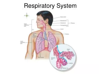
Respiratory (A&P, ABG’s, Assessment) Respiratory case studies Ron Pinkston, RRT Respiratory Therapist Staff Educator/ Team Leader
Indications for Oxygen Therapy • Hypoxemia • Excessive Work of Breathing • Excessive Work of Heart
Oxygen Delivery Devices • Oxygen Cylinder • Nasal Cannula • Simple Mask • Venturi Mask • Partial Rebreather • Non-Rebreather • Continuous Aerosol
Important points to consider! • Room air is 21% oxygen • The oxygen that comes out of the wall or oxygen tank is 100% oxygen • Whatever device or amount of flow we use to deliver the oxygen to the patient will determine the % of oxygen the patient actually inhales (FIO2)
24-40% FiO2 Nasal Cannula The nasal cannula supplies the patient with low to moderate concentrations of oxygen. Be sure to add humidifier bottle if the nasal cannula is run at > 3 lpm or upon patient request. Approximate FIO2 at a given liter flow: 1 lpm = 24% 2 lpm = 28% 3 lpm = 32% 4 lpm = 36% 5 lpm = 40% 6 lpm = 44%
Approximately 40-60% FiO2 Simple Mask The simple oxygen mask should be run between 6 and 10 lpm to ensure flushing and prevent CO2 accumulation in the face mask. This flow will result in an approximate FIO2 approximately 40 – 60%, depending upon the patient’s ventilatory pattern. Simple masks are most often used on the pediatric patient that does not tolerate a nasal cannula. Use caution when using O2 masks on nauseated patients, due to the increased risk of aspiration
Exactly 24-50% FiO2 Venturi Mask The Venturi mask (or “Venti” mask) delivers a specific FIO2 concentration (ranging between 0.24 & 0.50, depending on the adapter) to the patient using a recommended flow rate. Venturi masks are most often used for the COPD patient that requires an exact FIO2 with a higher flow than can be given with nasal cannula. When using the Venturi mask, be sure you do not block the jet port holes with the patient’s blanket or gown. This will effect the FIO2 being delivered. Each adapter is specified for FIO2 & flow
Approximately 60-80% FIO2 Partial Rebreather Mask The partial rebreather mask is often use when needing higher FIO2 in place of the nasal cannula to maintain a patient’s SpO2 > 90%. The partial rebreather mask delivers a moderate concentration of oxygen. (approximately 60 - 80%) Run the flowmeter at 8 - 15 lpmto ensure the reservoir bag does not deflate on inspiration to prevent CO2 buildup in the mask. Keep reservoir bag above patient’s blanket and make sure it is not twisted or kinked. Do not block intake ports. Provide alternate source of oxygen (nasal cannula) when patient is eating. Change mask whenever foreign material accumulates on the mask. No valves!
Approximately 80-100% FIO2 Non-Rebreather Mask The non-rebreather mask administers high concentrations of oxygen (approximately 80 - 100%) The non-rebreather is used for the patient without an artificial airway (ETT or tracheostomy), exhibiting severe respiratory distress or requiring increased O2 to keep SpO2 > 90%. Run the flowmeter at 8 - 15 lpm to ensure the reservoir bag does not deflate on inspiration to prevent CO2 accumulation in the face mask. Keep reservoir bag free from patient blanket and make sure it is not twisted or kinked. Do not block intake port one-way valves. Change mask whenever foreign material accumulates in the mask. Notice the 1-way valves!
Aerosol Devices Aerosol devices administer specific FIO2 concentrations (ranging from 28 to 100%, depending on dial setting) To provide an increased level of humidity at controlled oxygen concentrations. Liter flow is determined by the FIO2 dial setting
Oxygen Cylinder Remember ! • Cylinders must always be in appropriate carrier or cart, never left standing alone • Do not use if the cylinder gauge reads less than 500 psi • Return patient to flow meter on wall when transport is completed
The most common airway emergency is soft tissue upper airway obstruction. Complete upper airway obstruction is accompanied by marked inspiratory efforts without air movement. The first maneuver attempted should be the head-tilt, chin-lift maneuver. Airway Management Blocked airway
The oropharyngeal (oral) airway is designed for insertion along the tongue until the teeth or gingiva limit the insertion. The device lies between the posterior pharynx and the root of the tongue and thereby maintains a patent airway. The oral airway is adequate for comatose patients only and usually for limited periods of time Measurement of proper size Mouth to jaw line Oropharyngeal Airway
The nasopharyngeal (nasal) airway is a soft/pliable tube constructed so that it can be inserted through one of the nares and follow the posterior wall curvature of the nasopharynx and oropharynx. It is much better tolerated than the oropharyngel by the semicomatose and awake patient. Suctioning of the pharynx is possible with appropriately lubricated suction catheter. Sizing: Nose to ear lobe Nasopharyngeal Airway
Manual Resuscitator • Delivers high oxygen concentrations (90-100% FiO2) • Run oxygenflowmeter> 15 lpm • Position head with chin-lift/ head-tilt • Oral airway may be used • Position mask with narrow end over the bridge of the patient’s nose, ensuring tight seal Two-person One-person
Definitions Pulse Oximetry:measuring the estimated arterial oxygen saturation using a non-invasive method. SpO2:abbreviation for the estimated arterial oxygen saturation given as a percentage, via pulse oximetry. SaO2:abbreviation for the estimated arterial oxygen saturation given as a percentage, via arterial blood gas. Hypoxia:an abnormal condition in which the oxygen available to the body cells are inadequate to meet their metabolic needs Hypoxemia:abnormal deficiency of oxygen in the arterial blood.
How Do Pulse Oximeter’swork? Red and infrared light must pass through a perfused capillary bed of the finger, toe, ear lobe, neonate’s foot or hand. The light emitters must be positioned directly opposite of the photodector. The oximeter uses the ratio of Red / Infrared light that is absorbed by the hemoglobin to determine the oxygen saturation (Spo2).
Indications • daily monitoring • titration of supplemental oxygen. • special procedures (i.e. bronchoscopy, TEE, conscious sedation). • sleep studies. • ambulation To determine adequacy of oxygenation during
Clinical signs of hypoxia • Tachycardia • Arrhythmias • Tachypnea • Dyspnea • Restlessness • Cyanosis • Irritability • Decreased LOC
Non-invasive Easy to use Can be used continuously to monitor trends. Rapid response Inexpensive Must have good signal to be accurate Inaccurate under certain circumstances. Pressure sores/ burns from prolonged use. Pulse oximetry Advantages Disadvantages
pH • The relative concentration of hydrogen ion in arterial blood. • The chief component of acid-base balance. (acidosis)7.357.40 7.45 (alkalosis) “base”
PaCO2 is the partial pressure of carbon dioxide in the blood CO2 functions as a RESPIRATORYacid Normal value is 40 mmHg CO2 levels stimulate respiration PaCO2 pCO2(alkalosis)3540 45 (acidosis) “base”
PaO2 • Partial pressure of oxygen in arterial blood • Does not play an active role in acid/base balance • Normal for an adult < 60 years is 80-100mmHg • After the age of 60, normal PaO2 levels are slightly lower due to decreasing lung compliance Levels of Hypoxia Severe <40mmHg Moderate 40-60mmHg Mild 60-80mmHg Norm 80-100mmHg
Hypoxemia/ Hypoxia • Hypoxemia: Low oxygen content in arterial blood (seen on ABG) • Hypoxia: Inadequate oxygen at the tissue or cellular level (clinical judgment) • Clinical signs of hypoxemia • tachycardia • cyanosis • hyperventilation • restlessness, uncoordinated activities • hypertension
Bicarbonate (HCO3) • An indicator of the base reserve • Reflects the kidney and/ or METABOLIC function “base” HCO3(acidosis)2224 26 (alkalosis)
Oxygen Saturation (SaO2) • Percent of hemoglobin which is bound to oxygen (calculated) • Normal SaO2 is 95 - 100%
ABG Normal Values low end normal high end pH 7.35 7.40 7.45 pCO2 35 40 45 pO2 > 80 mmHg HCO3 22 24 26 BE -2 0 +2 acidic alkalotic alkalotic acidic acidic alkalotic alkalotic acidic
pH 7.33 PaCO2 64 HCO3 23 PaO2 65 Look at the pH is it normal (N)? is it acidotic (A)? is it a base (alkalotic) (B)? Assess Acid/ base balance (step 1) “A” “A” • Look at the pCO2 • is it normal (N)? • is it acidotic (A)? • is it a base (alkalotic) (B)? “N” • Look at the HCO3 • is it normal (N)? • is it acidotic (A)? • is it a base (alkalotic) (B)?
pH 7.33 PaCO2 64 HCO3 23 PaO2 65 Acidosis Acidosis Normal Match system to pH (step #2) “A” “A” “N” The acidosis is caused by the RESPIRATORY acids So this example is a Respiratory Acidosis
pH 7.33 PaCO2 41 HCO3 12 PaO2 65 “A” Acidosis “N” Normal “A” Acidosis Another Example The acidosis is caused by the METABOLIC acids So this example is aMetabolicAcidosis
COPD patients with chronic CO2 retention no longer breathe according to CO2 levels. These patients rely on the presence of hypoxemia (low PaO2) to stimulate respiration. Avoid giving too much O2 to these patients. Elevated oxygen levels may reduce their stimulation to breathe! Keep PaO2 between 50 - 60 (pulse-ox 85 - 90%). Baseline PaCO2 > 45 mmHg. Elevated HCO3 (to compensate for increased PaCO2) Baseline pH within normal limits. Chronic CO2 retention
Typical Baseline COPD: CO2 Retainer ABG’s on Room Air pH 7.40 PaCO2 50 HCO3 32 PaO2 55 “N” “A” “B” Moderate hypoxemia Compensated Respiratory Acidosis with moderate hypoxia
Admitting the Pediatric PatientAssessment of the Respiratory System Retractions • Intercostal • Suprasternal • Clavicular • Substernal • Subcostal
Admitting the Pediatric PatientAssessment of the Respiratory System • Respiratory Rate/Pattern • Do not rely on monitor to obtain accurate RR • Count for 1 full minute • Observe pattern (periods of apnea, paradoxical, rapid and deep) • Breath Sounds…Can be very tricky! • The infant or toddler will not remain motionless or quiet. • The infant or toddler will not take a deep breath upon command. • The room may be noisy. • The child or infant may become frightened and begin crying. • Listen during Insp. and Exp. Phase (2 cycles ideal) • Systematic Assessment; comparing segments from side to side
Hr 120-140 SpO2 85% RR 60 bpm Insp/Exp. Wheezing Inspiratory Crackles Paroxysmal cough Parents report periods of apnea Copious thick secretions Intercostal & Suprasternal Retractions Rhinitis Afebrile Normal WBC Crackles Wheezes Pulmonary Disorders in ChildrenCase #13 month old infant presents to pediatrics with:
Pulmonary Disorders in Children • CXR: Air trapping, atelectasis and infiltrates.
Pulmonary Disorders in ChildrenBronchiolitis • Management • Oxygen • Suction • IV hydration • Racemic Epinephrine Nebs • Bronchodilators • Antibiotics • Monitoring • Bronchiolitis Protocol (if ordered)
Pulmonary Disorders in ChildrenCase #2 • 10 month old presents to the ED. Mom states when she picked her son up from the daycare after work he was breathing harder and had trouble drinking his bottle. MD office sent her home with rx. For MDI and oral steroid. Mom states she is very worried; feels something is very wrong. -- HR 180 -- Suprasternal retractions • RR 80 -- SpO2 90% on 5 lpm HFH -- Frequent Coughing -- Nasal Flaring -- Febrile -- WBC increased -- Grunting -- RUL atelectasis • Tight exp. wheezing despite 1 hour continuous neb.