Download
1 / 51
510 likes | 537 Views
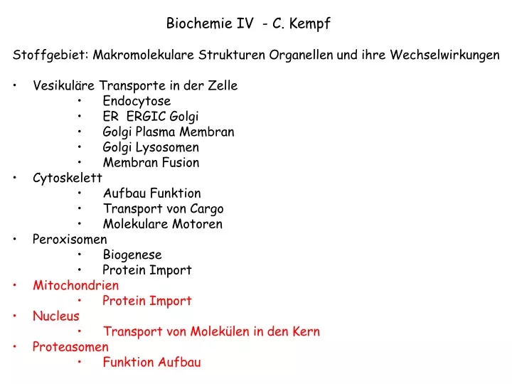
Biochemie IV - C. Kempf Stoffgebiet: Makromolekulare Strukturen Organellen und ihre Wechselwirkungen Vesikuläre Transporte in der Zelle Endocytose ER ERGIC Golgi Golgi Plasma Membran Golgi Lysosomen Membran Fusion Cytoskelett Aufbau Funktion Transport von Cargo Molekulare Motoren

E N D
Biochemie IV - C. Kempf • Stoffgebiet: Makromolekulare Strukturen Organellen und ihre Wechselwirkungen • Vesikuläre Transporte in der Zelle • Endocytose • ER ERGIC Golgi • Golgi Plasma Membran • Golgi Lysosomen • Membran Fusion • Cytoskelett • Aufbau Funktion • Transport von Cargo • Molekulare Motoren • Peroxisomen • Biogenese • Protein Import • Mitochondrien • Protein Import • Nucleus • Transport von Molekülen in den Kern • Proteasomen • Funktion Aufbau
Organelles specialized for ATP synthesis. • Most, but not all, proteins are encoded by the nuclear genome and synthesized in the cytosol. • Proteins must be transported to one of multiple membranes or compartments.
Import of the protein into the matrix is directed by an N-terminal signal sequence. • For polypeptides encoded by the nuclear genome, synthesis of the polypeptide is first completed in the cytosol. Transport occurs by a posttranslational mechanism. • Signal sequence at the N-terminus associates with the TOM complex located in the outer mitochondrial membrane. TOM is both a receptor for the signal sequence and a translocase. • The polypeptide is passed from TOM to TIM in the innermembrane. During the transport process, the polypeptide traverses both inner and outer membranes via the two translocators at a point known as a contact site. • The polypeptide is imported and the signal peptide is removed by a signal peptidase.
The energetics of import - import requires ATP hydrolysis and an electrochemical proton gradient. • ATP hydrolysis regulates the association of chaperone proteins in the cytosol that serve to keep the polypeptide in an unfolded state prior to association with TOM. • Electrochemical proton gradient in the inner membrane draws the signal sequence through TIM into the matrix. • Chaperone proteins in the matrix use the energy of ATP hydrolysis to pull the polypeptide into the matrix and guide proper folding.
The N-terminal signal sequence for mitochondrial import is a positively charged, amphipathic alpha helix. Hence, the membrane potential across the inner mitochondrial membrane may “electrophorese” the signal sequence through the TIM complex.
A second signal sequence in the polypeptide can be involved in directing a protein to a destination in the mitochondria other than the matrix. All of the signal sequences are contained within amino acid sequence of the imported protein, and they function subsequent to the functioning of the N-terminal signal sequence.
Transport into chloroplasts is similar to mitochondria except there is a 3rd membrane that can be targeted. Targeting the thylakoid membrane involves a second signal sequence. In the case of chloroplasts, the electrochemical proton gradient is at the thylakoid membrane where this gradient participates in transport. Transport across the chloroplast inner membrane is powered by GTP and ATP hydrolysis.
Roadmap of protein traffic. The genesis and function of internal compartments depends on the appropriate targeting of proteins Some of the green route illustrated.
Most newly synthesized proteins begin synthesis in the cytosol and their destinies depend on sorting signals encoded in the amino acid sequence of the protein. Proteins synthesized in the cytosol and lacking sorting signals remain in the cytosol. A small number of proteins do not begin synthesis in the cytosol. These proteins are found in mitochondria and chloroplasts. They are encoded by DNA present in these organelles and are synthesized by ribosomes residing in the lumen of these organelles.
A simple experiment shows that many sorting signals consist of a continuous stretch of amino acid sequence called a “signal sequence”. Fusing sorting signals to GFP is particularly good way to do this experiment.
Signal sequence Signal patch A more complicated sorting signal consists of a “signal patch” generated by the tertiary structure of the protein.
Nuclear envelope consists of two concentric lipid bilayers. • The perinuclear space is contiguous with the lumen of the ER. • Bidirectional transport occurs through the nuclear pore complexes.
The nuclear pore complex is an aqueous channel that allows diffusion of small molecules and proteins up to 60kD. Hence, transport of such molecules is passive.
Proteine grösser als 60 kD benötigen einen aktiven Transport. Wenn ein Protein aktiv in den Kern Transportiert wurde bleibt es dort ausser ein spezieller Mechanismus löst den Rücktransport durch die Kernpore aus. So genannter “Gated transport”. ?
Warum braucht es Import von Proteinen in Kern • Proteine, welche für die Funktionen im Kern gebraucht werden, werden im Cytoplasma synthetisiert. • Während der Zellteilung bricht der Kern auseinander. Kern und Cytoplasma werden gemischt. Diese Komponenten müssen in den Tochterzellen wieder sortiert werden. Laminine DNA Phosphorylierung der Laminine Fusion behüllter Chromosomen P P Interphase Kern P Fragmentierte Kern- membranen P Späte Telophase Prophase Frühe Telophase
In most cases, nuclear localization of large proteins relies on a signal sequence called a Nuclear localization signal or NLS. • NLS can be located anywhere in the primary sequence of the protein. • Usually arginine and lysine-rich and quite short. • There are some exceptions where nuclear localization relies on a signal patch.
The NLS directs the protein for transport through the nuclear pore complex, and proteins maintain their tertiary and quaternary structures during transport. When gold beads are coated with the NLS, the beads can be seen passing through nuclear pore complexes. The maximum size bead that can be transported is approx 25 (35-40) nm. Since the gold bead can’t compress, the opening of the pore must be able to expand.
Import Receptor Ran GDP Ran GTP Cargo Nuclear targeting Signal The NLS associates with soluble cytosolic proteins called nuclear import receptors. The nuclear import receptors also bind the nuclear pore complex so they serve to bring the protein containing the NLS to the nuclear pore complex.
Import Receptor Ran GDP Ran GTP Cargo Nuclear targeting Signal The protein-receptor complex moves through the pore but the mechanism of transport is not known.
Import Receptor Ran GDP Ran GTP Cargo Nuclear targeting Signal Once inside the nucleus, a protein called Ran-GTP displaces the cargo from the import receptor. Ran-GTP is a monomeric GTPase that binds and hydrolyzes GTP.
Import Receptor Ran GDP Ran GTP Cargo Nuclear targeting Signal The receptor must be recycled back to the cytoplasm for transport to continue. The Ran-GTP/receptor complex associates with the nuclear pore and travels out of the nucleus via the pore.
Import Receptor Ran GDP Ran GTP Cargo Nuclear targeting Signal When the Ran-GTP/receptor complex arrives in cytoplasm, Ran hydrolyzes the GTP to GDP and releases the receptor.
Export Receptor Ran GDP Ran GTP Cargo Nuclear Export Signal
Monomeric GTPases In the cell, this cycle only runs counter-clockwise because the cell maintains a high GTP/GDP ratio. The rate of the cycle is controlled by GAP and GEF because the intrinsic GTPase activity and rate of nucleotide exchange for the monomeric GTPase are slow. GAP and GEF lower the activation energies of these processes. • Monomeric GTPases are a family of proteins that exist in a GDP-bound state or a GTP-bound state. These two states have different activities. • The transition from the GTP state to the GDP state involves hydrolysis of the GTP and the rate of hydrolysis is accelerated by a GTPase-activating protein (GAP). • The transition from the GDP state to the GTP state is accelerated by a Guanine nucleotide exchange factor (GEF). • Note that one complete cycle of changes hydrolyzes GTP. This hydrolysis of GTP is used to power cellular functions.
An asymmetric distribution of a Ran-GAP and a Ran-GEF controls the activity of Ran in a way that allows Ran to mediate active transport through the nuclear pore complex. Ran-GAP in the cytosol causes Ran-GDP to predominate in the cytosol. Ran-GEF in the nucleus causes Ran-GTP to predominate in the nucleus.
Low concentration of Ran-GTP prevents Ran from disrupting the association of import receptor and NLS Ran-GAP Ran-GTP Ran-GDP H20 phosphate Cytosol perinuclear space perinuclear space Nucleus High concentration of Ran-GTP promotes dissociation of NLS from import receptor and promotes the export of NLS-free receptor. Ran-GEF Ran-GDP Ran-GTP GTP GDP
The nuclear import players: • Nuclear import receptor binds the cargo. • NLS is in the amino acid sequence of the cargo protein. • Ran-GTP and Ran-GDP are different forms of Ran bound either to GTP or GDP. Ran-GTP causes the NLS to dissociate from the Nuclear import receptor. • Ran-GAP is distinct from Ran but causes Ran to hydrolyze GTP. Hence, Ran-GAP promotes the conversion of Ran-GTP to Ran-GDP. • Ran-GEF is distinct from Ran but causes Ran to release GDP and bind a different molecule of GTP. Hence, Ran-GEF promotes the conversion of Ran-GDP to Ran-GTP.
Import process 1. Nuclear import receptor associates with cargo and brings the cargo to the nuclear pore. 2. Somehow the receptor/cargo complex moves through the pore. 3. Once in the nucleus, Ran-GTP displaces the cargo from the receptor. Ran-GTP is present in the nucleus because of the Ran-GEF. 4. Receptor/Ran-GTP exits the nucleus through the pore. 5. Once the receptor returns to the cytosol, Ran-GAP induces Ran to hydrolyze its bound GTP. 6. Ran-GDP dissociates from the receptor.
Export of RNA and proteins relies on nuclear export receptors associating with nuclear export signals (NES) found on proteins and RNA-bound proteins. • Nuclear export receptors are structurally similar to nuclear import receptors. • Nuclear export receptor bind the nuclear export signals and bring the protein to the nuclear pore complex for subsequent transport. • Transport occurs through the same pores through which proteins are imported from the cytosol. • Ran regulates the interaction between the export receptor and the “NES”. The Ran-GTP promotes association of the receptor/cargo complex with the pore in the nucleus and hydrolysis of the GTP on the cytosolic side causes the resulting Ran GDP to dissociate the export receptor from its cargo. • Note - nuclear export receptors do not bind directly to RNA, they bind proteins bound to the RNA.
Protein Abbau ist essentiell für die Zelle... • Bereitstellen von AS für neue Pproteinsynthese • Entfernen überschüssiger Enzyme • Entfernen von Transcriptions Factorens (gene action), welche nicht mehr benötigt werden
Es gibt zwei hauptsöchliche intrazelluläre Organellen, welche defekte und nicht mehr benötigte Proteine abbauen 1)Lysosomen : Abbau extrazellulärer Proteine, welche von der Zelle aufgenommen werden (Endozytose, Phagozytose) und 2)Proteasomen: Abbau endogener Proteine; z.B. Transkriptionsfaktoren, Cycline faslch gefaltete Proteine, virus codierte Proteine, defekte proteine….. [cystic fibrosis beruht auf einem beschleunigten Abbau des Cl transporters] Proteasomen bauen Proteine zu Peptiden ab, welche nachfolgend durch Exopeptidasen hydrolisiert werden
PROTEASOM • Ein Protein verdauendes „Organell“? • Eine Enzymkomplex für nicht-lysosomalen Protein Abbau • PROTEOSOMEN grosse multi Enzym Komplexe Eine menschliche Zelle enthält zwischen 20,000 - 30,000 Proteasomen. • Das Proteasom ist ein Zylinder förmiger Komplex bestehend aus 4 Teilen (MW 2‘400 kD) • „Deckel“ aus 9 Proteinen • Regulatorischer Teil welcher nur ubiquinierte Proteine akzeptiert • „abbauender Teil“ (4 protein Ringe“ enthaltend die Protease Aktivitäten • Basis Teil
Die Blockierung des proteasomalen Abbaus zellulärer Proteine kann eine Zelle auch schützen: • Die Blockierung der Proteasomen durch Inhibitoren (MG132, Aclarubicin…) führt zu einer Akkumulierung von ubiquitinierten defeketen Proteinen. Dies wiederum löst eine HEAT-SHOCKReaktion aus, welche die zelle vor toxischen Substanzen oder erhöhten Temperaturen schützen kann • Pharma Konzerne forschen z.Z an Preoteasom Inhibitoren mit dem Ziel Medi zu entwickeln um: • Zellen vor „Ischämie“ zu schützen • Organe nach Transplantationen zu schützen • die lebensdauer von Zyklinen und Transkriptionsfaktioren zu beeinflussen • Krebstherapie z.B. Velcade
Protein Abbau durch Proteasomen... Ist abhängig von Ubiquitin. Proteine welche durch Proteasomen abgebaut werden sollen müssen ubiquitiniert werden (Erkennungssignal) Ubiquitin: globuläres Protein, 76 aa (praktisch identische aa Sequenz in Bakterien, Hefe oder Säugern) MQIFVKTLTG KTIALEVESS DTIENVKAKI QDKEGIPPDQ QRLIFAGKQL EDGRTLADYNIQKESTLHLV LRLRGG (K Anhängen weiterer Ub‘s; G bindung an „Zielprotein“) Ubiquitin Ligase Enzyme [ E1, E2, E3 ] koppeln Ubiquitin an die Proteine
Ubiquitinierung von Proteinen erfolgt durch ein komplexes System bestehend aus • Ub aktivierendem Enzym (E1) • UB konjugierendem Enzym (E2) • Substrat erkennendes Protein ( E3)
O C O C = = E3 ATP PPi Lys48 O C E1 E1 = HS- COOH -S- E2 E2 HS- -S- ε-NH2 -S Cys
O C O C O C- O C- O C- = = = = = O C E2 = -S - E3 E3 E3 ε-NH2 -S Cys O C- = ε-NH ε-NH -S HS Cys Cys n
Die „zentrale Einheit enthält 3 Protease Aktivitäten: Trypisn ähnliche Protease Chymotrypsin ähnliche Protease Peptidyl-glutamyl peptide hydrolase Diese Enzyme spalten das ubiquitinierte Protein in Peptide, welche dann von cytosolischen Proteasen weiter verdaut werden. Ubiquitin wird rezykliert. ATP ADP
electron density micrograph of the 26S proteasome The 20S proteasome is a hollow cylinder structure composed of 4 stacked rings, each containing seven subunits. The outer rings consist of seven different alpha-type subunits whereas the inner rings are composed of seven different beta-type subunits. The channel spanning the centre of the 20S proteasome is divided in three cavities. Both outer cavities are located at the interphase of alpha- and beta-rings. The central cavity containing the proteolytically active sites is build up from beta-rings.
Aktive Stellen im Hefe 20S Proteasome Chymo PGPH Tryp Tryp PGPH Chymo
Regulatorischer Teil (19S): • in Hefe besteht dieser regulatorische Teil (RP) aus min 18 Untereinheiten • 6 (Rpt 1-6) sind AAA ATPase • 12 Rpn
Loklaisierung der Proteasomen: • Cytoplasma • Kern • ER assoziert
Hypothetisches Model der Proteasome ER Assoziation/Funktion Cytoplasma ER Lumen sec61 Ub conjugating enzyme ? Cytoplasma
Cytoplasma TAP Transporter „Protein ?“ Class I MHC ER Lumen