Download

1 / 13
130 likes | 244 Views
Imaging tests are very important for breast cancer Diagnosis. If there is a suspicious area, these tests help find out whether it is cancerous and also determine how far it has spread

E N D
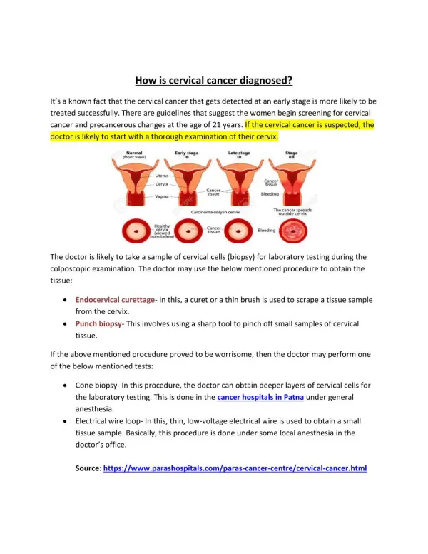
Breast Cancer Can Be Diagnosed Lump or thickening of breast often painless Change in the size of breast Discharge of bleeding from nipple Discoloration of the skin on breast Inversion of the nipple Change in color or appearance of areola Change in shape of breast Dimpling on the skin
Common Kinds Of Breast Cancer • Ductal carcinoma • Ductal carcinoma in situ (DCIS) • Invasive ductal carcinoma • Lobular carcinoma • Lobular carcinoma in situ (LCIS) • Invasive lobular carcinoma
Ductal carcinoma – A type of tumor primarily present in ducts of a gland. • Ductal carcinoma in situ - an uncontrolled growth of cells within the breast ducts. “in situ” means “in its original place”. It is considered as stage 0 cancer , it is curable. • Invasive ductal carcinoma - it begins in the milk ducts of the breast. It grows through the duct walls and into the surrounding breast tissue and also spreads to other areas of the body.
Lobular Carcinoma – lobulas are the glands that produces milk. The milk then moves to the ducts. Majority of the breast cancer starts in the ducts some through lobulas. Lobular Carcinoma In Situ (LCIS) – Abnormal growth of cells in milk producing glands. It is commonly diagnosed after biopsy. • Invasive Lobular Carcinoma – Condition begins in one of the breast lobules and then spreads to other parts of the breast. Feel of a thickened area or a hardening in part often occurs in the area above nipple moving in the direction of armpit.
Imaging Tests Imaging tests are very important for breast cancerDiagnosis. If there is a suspicious area, these tests help find out whether it is cancerous and also determine how far it has spread. Doctors may use several imaging tests such as diagnostic mammography, ultrasound and MRI scans.
Diagnostic Mammographyis used to take pictures of the area concerned and is usually used when a woman experiences nipple discharge, finds a lump or detects an abnormal result on her screeningmammogram
Ultrasoundis used to help distinguish between a solid cancerous mass and a fluid-filled cyst. It can also be used to locate the position of the known tumor.
MRI (Magnetic Resonance Imaging) is done after the cancer has been diagnosed. MRI scans are used to check the other breast for cancer.
Biopsya small amount of tissue is removed from the breast area and examined under a microscope. The reason why a biopsy is so important is because even though other tests may help in detecting cancer, only a biopsy can make a proper diagnosis.
Apart from these standard tests, the doctor may also recommend additional tests which are generally prescribed for patients with more advanced stage disease. Tests such as x-rays, to look for cancer that has spread from the breast to the lungs, bone scans, used to look for spread of cancer to the bones, CT or CAT (computerized tomography scan) scans, used to look for distant tumors and PET (positron emission tomography) scans, used to check the extent to which cancer has spread are some of the additional tests done.
Stages of Breast Cancer • Stage I And II Breast Cancer refers to a tumor less than 2 cm in size that is node-negative. Stage II tumors are those with spread to the axillary lymph nodes or a tumor size larger than 2 cm but not larger than 5 cm • Stage III Breast Cancer these consist of large breast tumors (greater than 5 cm across), those with extensive axillary nodal involvement, or nodal involvement of the soft tissues above or below the collarbone • Stage IV Breast Cancer — Stage IV breast cancer refers to tumors that have metastasized to areas outside the breast and lymph nodes to the brain, bones, skin, or other organs. The primary tumor may be any size, and there may be any number of affected lymph nodes. This is referred to as metastatic breast cancer