Download
1 / 20
200 likes | 451 Views
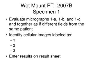
Wet Mount PT: 2007B Specimen 1. Evaluate micrographs 1-a, 1-b, and 1-c and together as if different fields from the same patient Identify cellular images labeled as: 1 2 3 Enter results on result sheet. Micrograph 1-a High Power. 3. 1. 2. 2. 3. 1. Micrograph 1-b High Power. 1. 3.

E N D
Wet Mount PT: 2007BSpecimen 1 • Evaluate micrographs 1-a, 1-b, and 1-c and together as if different fields from the same patient • Identify cellular images labeled as: • 1 • 2 • 3 • Enter results on result sheet
Micrograph 1-a High Power 3 1 2 2 3 1
Micrograph 1-b High Power 1 3 2 2
Micrograph 1-c High Power 2 1 1 3
Wet Mount PT: 2007B Specimen 2 • Evaluate micrographs 2-a, 2-b, and 2-c together as if different fields from the same patient • Identify cellular images labeled as: • 4 • 5 • 6 • Enter results on result sheet
Micrograph 2-a Low Power 5 5 4 4
Micrograph 2-b Low Power 4 4 6 5 4
Micrograph 2-c High Power 4 6 4 5
Wet Mount PT: 2007B Specimen 3 • Evaluate micrographs 3-a, 3-b, and 3-c together as if different fields from the same patient • Identify cellular images labeled as: • 7 • 8 • 9 • Enter results on result sheet
8 7 9 Micrograph 3-a Low power
8 7 7 8 Micrograph 3-b Low Power
Micrograph 3-c High Power 9 9 9
Wet Mount PT: 2007B Educational Challenge • Evaluate the following micrographs • These will not be graded!
Phase Contrast – High Power Identify this cell
1a – Healthy Trichomonas 1b – Dying Trichomonas (rounded up) For size reference: 2=Squamous Epi 3 = Yeast cell
Trichomonas vaginalis Flagella Nucleus Flagella Nucleus
Trichomonas • Trichomonas are parasitic protozoa. • They can be very motile • When they begin to die (within 10 minutes of specimen collection), they become sedentary and begin to round up. • Cells also fatten when grown in culture medium (as in the previous slides). • Trichomonas should only be reported when motility is observed
Trichomonas have a very complex structure. They have four flagella facing ‘forward’ and a fifth facing ‘backward’ which is attached to an undulating membrane. Sometimes, an ‘axiostyle’ or structural shaft can be seen – often it can’t The cells are oval in shape, 10-23 µm in length
Assessment of Cell Size • Use nucleus of Squamous Epi Cell as size marker • Nucleus = 15 microns • Yeast = 5-7 microns • RBC = 6-8 microns • WBC = 15 microns • Trichomonas = 20 microns