
Vision Loss and Blind Spots in Eye Health
E N D
Presentation Transcript
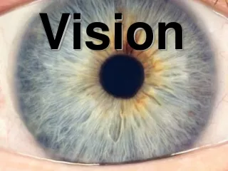
Vision • Vision is much more complicated because these signals have to be processed into a 3-D image
Fundus Photograph – Hu Eye Fovea Optic Disk
Blind Spot • Find Your “blind spot” Optic Disk • Cover 1 eye with the hand from the same side of your body, i.e., L hand over L eye • Fully Extend your free hand with your index finger pointing upward directly in front of your other eye. Note what ever object your index finger is covering and move your finger just out of the way so you can fix your sight on that object. • Now wiggle your index finger while slowly moving it to the right, left up and down within your peripheral view . • Just to the lateral periphery of your fixed view, you will notice you are “blind” to your wiggling finger. • Do the same for your other eye. • Where is your blind spot for your other eye? • How come you are never aware of your blind spot?
Amsler Grid Normal Pre-convergence Appears Normal to Patient but ….. Crowding, Completion and Fixation “Hide” the Details Vision Loss
Completion The classic “Kaniza Triangle” – one can clearly see a triangle, even though the borders are composed of virtuallines formed by the brain
Fixation – 1 moment please When viewing this face, we automatically scan it with our fovea to form a complete, clear image. The Amsler grid cannot force fixation on acentral point, thereby it cannot prevent the peripheral scanningwhich may mask a non-foveal defect. This is the actual track of an eye looking at this picture, even though the observer was instructed to stare at a single point on the face
+ S R Z + R Crowding While staring at the yellow cross in the upper box, try to read the middle letter on the right. Now try to do the same thing with the lower box. It is easier with the lower box where the “R” stands alone. The neighboring lines in the Amsler Grid reduce the patient’s ability to isolate and detect visual distortions.
Can you see which are “popping / pimpling out” of the page and which are “pressing” dimpling / indenting in to the page?
Can you see which are “popping / pimpling out” of the page and which are “pressing” dimpling / indenting in to the page? Does it make a difference in this view?